Natural remedies for an overactive bladder

An overactive bladder (OAB) is a condition where you are not able to hold urine in your bladder due to involuntary bladder muscle contractions.  It causes a frequent, sudden and difficult-to-control urge to pass urine, which may lead to unintentional loss of urine. You may also feel the urge to pass urine 8 or more times in 24 hours.

With an overactive bladder, the first-line treatment is usually behavioral and lifestyle changes. But if the condition remains troublesome even with the changes, further treatment may include medications, bladder injections, nerve stimulation, percutaneous tibial nerve stimulation (PTNS), or surgery. 

So what are the natural remedies for an overactive bladder?

  1. Eliminating bladder irritants

Symptoms of an overactive bladder can be relieved by avoiding foods and drinks known to irritate the bladder. 

Common culprits include alcohol, caffeine, artificial sweeteners, citrus fruits and juices, and chocolate. Other foods and drinks to avoid or reduce are sodas and fizzy drinks, cranberry juice, corn syrup, tomatoes, sugar, honey, vinegar, dairy, and spicy foods.

Since irritation from foods and drinks vary from person to person, you can benefit by keeping a diary of what you take and the associated bladder symptoms. A diary can help you work out which foods are causing you problems.

  1. Making lifestyle changes 

Quitting smoking can remedy an overactive bladder. That’s because smoking tends to make the symptoms of the condition worse, while the coughing fits associated with smoking tend to increase OAB-associated leakage of urine

Another change that can improve an overactive bladder is withdrawal of medications contributing to the problem. For instance, diuretics, antihistamines, muscle relaxants, sedatives, narcotics, and alpha-adrenergic antagonists worsen OAB symptoms. So if you’re taking any medications you suspect to be behind the problem, speak with your doctor about alternatives. 

Also, losing weight can reduce the pressure on your bladder and pelvic floor muscles, and improve your bladder control.

  1. Using bladder control techniques

There are a number of bladder control techniques you can apply, including scheduled urination, delayed urination, and double-void technique. 

    • Scheduled urination

By scheduled urination, you keep a diary of your urinary habits, such as bathroom visits, any leakages, and urgency symptoms. Based on the patterns in your diary, you start scheduled bathroom trips aimed at adding at least 15 minutes to your normal urination times. 

For example, if you pass urine every 60 minutes, you change this and schedule your bathroom breaks for every 75 minutes. Then you stick to these scheduled visits regardless of whether you feel the urge to pass urine or not. After that, you gradually increase the time interval between your bathroom visits 

    • Delayed urination

Delayed urination means every time you feel the urge to go, you try to delay by some minutes, if possible, usually by at least 5 minutes. You can use relaxation techniques, such as deep breathing to help delay. The aim is to gradually increase the delay time until you have achieved a 3-4 hour gap between your bathroom visits.

    • Double voiding

Double voiding technique is useful if you feel your bladder does not empty fully. It is also worthwhile to double void before bedtime. 

To do so, you sit in the toilet, lean slightly forward, rest your hands on your knees or thighs, and urinate as normal. Then you remain in the toilet and wait for another 30 seconds, leaning slightly further forward to urinate once more.

  1. Doing kegel exercises

Kegels involve strengthening exercises on the pelvic floor muscles, which are the muscles that help to control urine flow. To locate your pelvic floor muscles, try to stop urine flow midstream. If you succeed, it means you have located the muscles.

Now, practice squeezing these muscles for 10 seconds and relaxing for 3 seconds, repeating the pattern 10 times. Then try to do 3 sets of 10 repetitions daily. Deep breathe to make the process easier.

  1. Managing your fluid intake

Drinking plenty of water is essential for your health, and you shouldn’t stop because of an overactive bladder. 

That’s because taking too little water allows your urine to be more concentrated, which may irritate your bladder lining and increase urgency. But excess fluid intake may also make frequency symptoms worse, such as fluid intake before bed contributing to urinating during the night.  

Make sure to drink 6-8 glasses of water daily, but to avoid liquid intake 2-3 hours before going to bed.

  1. Using herbs and supplements

There are a number of herbs and supplements that are effective in treating an overactive bladder. Speak with your urologist about these options for recommendations, but don’t use them without your doctor’s advice. 

Effective herbs and supplements include:

  1. Gosha-jinki-gan

This is a blend of 10 traditional herbs that help with overactive bladder by increasing the bladder’s capacity and lowering bladder contractions. It reduces frequency, urgency, and nighttime urination in people with the condition. 

  1. Capsaicin

Capsaicin is an extract from chili peppers. It works by blocking signals from the nerves in the bladder and ensures the bladder holds urine longer. Plus, it helps to manage pelvic pain syndrome, of which overactive bladder is usually a symptom.

  1. Pumpkin seed extract

The extract is effective in improving pelvic floor health. It helps to strengthen pelvic floor muscles and enhances control of muscles involved in passing urine.

  1. Magnesium hydroxide

Magnesium helps maintain normal blood pressure, reduces muscles spasms, and allows the bladder to empty fully. As an alternative to magnesium hydroxide, you can add magnesium rich foods, like bananas, kale, pumpkin seeds, and cashews to your diet if your doctor deems it safe.

  1. Saw palmetto

Saw palmetto has been tested in men with an enlarged prostate, where it has demonstrated efficacy in treating the condition as well as relieving bladder control symptoms. Hence, using it helps improve bladder issues associated with OAB.

  1. Resiniferatoxin

This is a chemical derived from a cactus-like plant. It is effective in blocking signals from the bladder nerves to the brain; hence, reducing the strength of the urge to pass urine, allows the bladder to hold urine longer, and relieves symptoms of an overactive bladder.

  1. Vitamin D

Increased intake of vitamin D lowers the risk of pelvic floor disorders, such as bladder leakage, especially in women. For older adults, more vitamin D in the diet or as a supplement effectively prevents bladder leakage.

  1. Bromelain

This is an enzyme found in pineapple plant. It has powerful anti-inflammatory abilities and prevents bladder irritation. Therefore, it has a positive effect on those with an overactive bladder.

  1. Cornsilk

Cornsilk refers to the hair-like threads found on top of the ears of corn and beneath cornhusks. It is rich in flavonoids and is helpful in relieving bladder control symptoms by reducing inflammation and protecting the bladder wall against irritation.

  1. Cleavers

This is a wild flower with tiny hooks on its stems, seeds, and leaves. It reduces bladder inflammation and protects the bladder wall from irritation.

At Advanced Urology Institute, we are known for our comprehensive, accurate diagnosis and tailored approach to treating urological disorders. With an overactive bladder, we make sure to establish the underlying cause, whether weak or stretched pelvic floor muscles, an enlarged prostate, chronic UTI, obesity, diabetes, or a nerve problem. 

Then, as first-line treatment, we often recommend natural remedies such as behavioral, lifestyle and dietary changes. For a more bothersome and persistent overactive bladder, we offer a variety of surgical and non-surgical interventions.

For more information on an overactive bladder and other urological disorders, visit the site “Advanced Urology Institute.”

7 Signs You Might Have Kidney Stones

An elevated amount of salts and minerals in urine can cause kidney stones. Also called renal calculi, kidney stones are hard deposits made of crystal-forming substances such as calcium, uric acid and oxalate, found in urine.  The stones vary in size, with some being too small, some a few inches across, and others large enough to take up an entire kidney. 

For smaller renal stones, there are usually no associated signs. The stones can travel from the kidneys through ureters, bladder and urethra without causing any problems.  And drinking plenty of water really helps in passing these stones. 

But for larger kidney stones, signs appear as the stones move from one part of the urinary tract to another.  An example is when a moderate or large stone moves from the kidney to the ureter. It immediately causes obstruction and produces agonizing pain. Such stones require a procedure, such as shock wave lithotripsy, to break up and remove them. 

So what are the signs that you might have kidney stones?

  1. Pain in your lower back, side, or belly

Kidney stones produce one of the most severe types of pain—comparable only to getting stabbed by a knife or pain during childbirth. The pain tends to begin when the stone moves into the ureter, causing a blockage and pressure buildup in the kidney. This pressure activates nerve fibers to send pain signals to the brain.

The pain starts suddenly and changes location and intensity as the stone moves. It also comes and goes in waves, with each wave lasting a few minutes, disappearing, and then coming back. The pain usually occurs along the side and back, below the ribs, but can radiate to the belly and groin area as the stone moves down the urinary tract.

  1. Tossing and turning

The sudden episodes of kidney stone pain last 20-60 minutes. But they are so severe that they don’t allow you to sit still. So you’re forced to move around, toss and turn, in order to find a more comfortable position.

  1. Burning sensation during urination

When a kidney stone reaches the junction between the ureter and bladder, it causes sharp or burning pain during urination. It is quite easy to mistake the stone for a urinary tract infection (UTI).  Of course, it is also common to have an infection alongside a kidney stone.

Apart from pain during urination, kidney stones can cause urinary frequency and urgency as they pass to the lower part of the urinary tract. That’s because a stone irritates the walls of the bladder and causes contraction, resulting in the urge to pass urine. You may find yourself running to the bathroom frequently or feeling the urge to go throughout the day and night.

  1. Nausea and vomiting

Kidney stones can cause obstruction of urine flow. This makes urine to back up, stretching or swelling the kidneys.  Eventually, this may lead to nausea and vomiting. 

Also, due to the excruciating pain associated with kidney stones, you may experience nausea and vomiting as one of the responses.

Equally, due to the sharing of nerve connections between the kidneys and the gastrointestinal tract, the presence of stones in the urinary tract disrupts nerves in the intestinal tract, resulting in stomach upset. 

  1. Blood in urine

Kidney stones irritate the delicate tissues that line the urinary tract, including inside the ureter. As a result, there may be significant, microscopic, or moderate bleeding, which results in blood in urine (hematuria). 

So as a sign of kidney stones, your urine may look grossly red, pink, or brown. You may also have blood in urine, but in quantities that are too small to notice with the naked eye. In that case, a urine test may be necessary to detect the urine.

  1. Fever and chills

Though fever is not a common sign of kidney stones, it may occur when the stones block urine flow or if the stones cause conditions that allow for an infection.  Like fever, chills tend to occur due to an infection that arises as a complication of kidney stones.

When they occur, fever and chills are usually a medical emergency. And so, the obstruction should immediately be dealt with through a procedure such as shock wave lithotripsy to enable antibiotics to pass through the obstructed area.

  1. Smelly or cloudy urine

Urine that is healthy tends to be clear and without a strong odor, but turbid, smelly urine might indicate an infection. So, while foul-smelling or cloudy urine does not directly indicate kidney stones, it may point towards an infection that arises as a complication of renal stones.

Generally, more than 16-percent of people with acute kidney stones tend to have UTIs. And whether it occurs with or without fever, the combined presence of a UTI and kidney stones is a surgical emergency.

When should you see a doctor?

You should see your doctor when you have agonizing pain, nausea, vomiting, bloody, turbid or smelly urine, fever or chills. It is advisable to seek immediate medical attention when you have pain that is so severe that you can’t get comfortable. For more information on kidney stone treatment, visit the site “Advanced Urology Institute.”

How long does radiation treatment take?

Key takeaways

  • Radiotherapy is a cancer treatment that uses high doses of radiation to destroy cancerous tumor cells. It can be administered externally or internally, and the length of treatment depends on the type, location, and characteristics of the tumor.
  • External radiotherapy is typically given daily, from Monday to Friday, for five to eight weeks, with breaks on the weekends to allow healthy cells to recover. The length of treatment may vary depending on the specific tumor and the location.
  • External radiotherapy is a painless process that typically takes 30-45 minutes, including set-up time, but the actual treatment only takes 2-5 minutes. The patient lies on a treatment table and is positioned under the radiation machine while special shields or blocks are used to protect healthy tissues.

Radiation therapy delivers controlled, safe and effective doses of radiation to cancerous tumors. The tumor cells are exposed to high doses of radiation that destroy their genetic material and eventually damage or kill them. Hence the cancer can no longer grow, multiply or spread after the treatment. Though the radiation affects all the cells, healthy ones are able to recover fully from the effects of the treatment.

Radiotherapy is generally administered either externally or internally. During external radiation therapy—the most common form of the treatment—a machine is used to direct high-energy rays at the cancer. In contrast, internal radiotherapy (also called brachytherapy) uses a radioactive source that is temporarily or permanently implanted directly into the cancerous area.

So how long does the radiation therapy take?

The exact duration of the treatment depends on the type, characteristics, and location of the tumor.  The length of treatment also depends on the dosage to be delivered, the number of fractions to be given, the treatment plan created by the radiation oncologist, and whether it is external or internal radiotherapy.

For example, if you have a deeper tumor, then you may require a more-focused beam delivered for a shorter period of time. But if you have a larger, shallow tumor, then you may need treatment for a longer period.

Equally, since the radiation must be given in a way that has minimal adverse effects on healthy cells, a shorter length of exposure is necessary if the tumor is located in more delicate organ (like the brain) or is in close proximity to very sensitive body tissues.

Your radiation oncologist will assess your tumor and make the necessary prescription. Then, by working with your radiation oncology team, the oncologist will determine how best to deliver the prescribed dosage, how many treatments are necessary, and how long it should take.

Next, your radiation oncologist will oversee the simulation of treatment to ensure that appropriate dose is given to the right location and that as little as possible reaches normal tissue. The simulation is followed by the testing of the delivery set-up to check the performance and positioning of the equipment to be used to give the treatment. After that, your treatment visits begin with the radiation therapist in charge of administering the daily fractions.

How long does external radiation therapy take?

External radiotherapy is typically delivered daily, from Monday through Friday, for five to eight weeks. Weekend breaks are factored into the treatment schedule to allow healthy cells to recover. Each treatment is offered on an outpatient basis.

But there are exceptions to this schedule.  Some tumors may require treatment for less than five days per week and only need treatment for one to two weeks. Specifically, shorter durations of two or three weeks in length are commonly used in palliative care—the use of radiation to relieve cancer symptoms. Still other tumors, such as certain brain cancers, may require just a single treatment.

External radiation therapy is a painless process and is almost like having a regular x-ray. You’ll be in a room for 30-45 minutes because of the time it takes to set up equipment and place you in the correct position, but the actual treatment takes 2-5 minutes.

Once in the room, you’ll lie on the treatment table. You’ll then be positioned under the radiation machine. Your radiation therapist will place special shields or blocks between the machine and other parts of your body to protect normal tissues. Thereafter, you’re expected to remain still, though you don’t have to hold your breath.

After you’re in the correct position, your radiation therapist will move into a separate, nearby room to turn on the machine and begin the actual treatment. The therapist will watch you on a monitor and you’ll be able to communicate with the therapist through an intercom.

How long does internal radiation therapy take?

Brachytherapy uses radiation implanted inside the body to treat cancer. The radiation implant is placed as close as possible to the tumor in order to concentrate the radiation on the cancer cells and minimize radiation damage on normal tissue around the tumor.  The radioactive material is sealed in a thin wire or hollow tube (catheter) and implanted directly into the cancer affected area on a temporary or permanent basis.

Internal radiation therapy is used when the oncologist decides that the best way to treat the tumor is to expose it to a higher radiation dose. The radioactive implant is closer to the cancerous cells and delivers a higher dose over a shorter period of time. The treatment is ideal for several types of cancers, including breast cancer, brain tumor, gynecological cancer (like ovarian and cervical cancer), lung cancer, and head and neck cancer.

The time taken for brachytherapy and whether it is done on an inpatient or outpatient basis depends on the type of therapy used and the nature of the cancer. In some cases, internal radiotherapy can be completed within three to five outpatient treatments of a few minutes each. But in other cases, the radioactive implant may be left in place for up to a week and there is need for a hospital stay during that period.

Finishing your radiation therapy sessions

It is important to finish all sessions of radiotherapy. And never to miss or delay treatments because this may reduce the effectiveness of the radiation in killing the tumor cells.

You should remember that your doctors are not just bothering you with making several hospital visits. There is need for different sessions because radiation is destructive and should not be delivered all at ago. In fact, if the recommended dosage was to be given once, it would pose a greater risk to healthy tissues and produce more adverse effects.

At Advanced Urology Institute, we spend the necessary time, effort and expertise to design a detailed treatment plan for radiation therapy. We offer the treatment in conjunction with other therapies and with the help of experienced oncologists and a multidisciplinary cancer care team.

We are also committed to reducing the time our patients take in treatment and often implement maximized aggressive treatments when necessary. Contact us today to learn whether radiotherapy is right for you and the various options available for you. For more information, visit the site “Advanced Urology Institute.”

References

What can cause an elevated PSA?

Key takeaways

  • The PSA test measures the quantity of a protein produced by cells of the prostate gland in a blood sample. It is typically used to screen for and monitor prostate cancer in men.
  • Elevated PSA levels can also be caused by non-cancerous conditions such as age, prostatitis, benign prostatic hyperplasia (BPH), and urinary tract infections.
  • To use an elevated PSA as the basis for ordering a prostate biopsy, it is now recommended that the level of PSA is monitored over time and any changes are monitored regularly, with a suspicious lump detected during a DRE being a more accurate basis for suspecting prostate cancer.

Prostate-specific antigen (PSA) is a protein produced by cells of the prostate gland. It is synthesized by both normal and malignant cells and released in blood. The PSA test measures the quantity of this protein in a blood sample, which is then reported in nanograms of PSA per milliliter (ng/mL) of blood. A PSA level of 4.0 ng/mL and below is often considered normal.

What causes an elevated PSA level?

The blood PSA level is typically elevated in men with prostate cancer. Therefore, the test is usually ordered in conjunction with the digital rectal exam (DRE) to screen men that are asymptomatic for prostate cancer. It is also recommended for monitoring the progression of prostate cancer in men already diagnosed with the disease, and to test men with prostate symptoms to find out the nature of their problem.

Apart from prostate cancer, there are a number of conditions that may increase the PSA level. For instance, PSA is elevated with age, usually due to enlargement of prostate tissue over the years. Prostatitis (inflammation of the prostate), which is a condition common in men under 50 years due to bacterial infection, tends to result in increased PSA level. Other conditions that lead to increased PSA level include benign prostatic hyperplasia (BPH), urinary tract infections, prostate injury, recent ejaculation, high parathyroid hormone, and surgical procedures.

Normal versus abnormal PSA level

Although a PSA level of 4.0 ng/mL or below is often considered normal, the level of the protein can vary over time in the blood of the same man, making what is usually taken as the normal range less accurate. In fact, studies have indicated that some men with PSA level below 4.0 ng/mL have prostate cancer while many men with levels above 4.0 ng/mL have been found free of the cancer.

Besides, due to the various factors that may cause a fluctuation of PSA level, such as age, prostatitis, BPH, and urinary tract infections, having a fixed normal range for all men is unreliable in some cases. Equally, since PSA test results vary from one laboratory to another and because drugs like Dutasteride (Avodart) and Finasteride (Proscar) that are used to treat BPH tend to lower PSA level, a single elevated PSA may not be very helpful.

Therefore, to use an elevated PSA as the basis for ordering a prostate biopsy to ascertain whether prostate cancer is present, it is now recommended that the level of PSA is monitored over time. A continuous trend of increasing PSA in blood over a prolonged period of time, together with a suspicious lump detected via the DRE, is a more accurate basis for suspecting prostate cancer and ordering for a prostate biopsy.

Elevated PSA in prostate cancer screening

For men without symptoms of prostate cancer, an elevated PSA level may be followed by a repeat PSA test to confirm the original finding. And if the PSA level is still high, the urologist may recommend that more PSA tests and digital rectal exams be done at regular intervals so that any changes can be monitored over time. If the PSA level continues to rise or if a suspicious lump is found during a digital rectal exam, the doctor may now order for confirmatory tests.

For example, a urine test may be requested to establish if the rising PSA level is due to a urinary tract infection. Likewise, imaging tests like cystoscopy, x-rays or transrectal ultrasound may help to show the size and nature of any lump.

And if the tests show there could be prostate cancer, the urologist will recommend a prostate biopsy.  Multiple samples of prostate tissue are collected by inserting hollow needles into the prostate through the wall of the rectum. The samples are examined by a pathologist to confirm whether the cells are cancerous or not.

Elevated PSA in monitoring prostate cancer treatment

After treatment for prostate cancer, the urologist will want to continue to monitor the PSA level to establish whether the disease is recurring or not. An elevated PSA level after treatment is usually the first sign that the cancer is recurring. In fact, an elevated PSA after treatment often happens many months or years before the signs and symptoms of prostate cancer recurrence show.

A single elevated PSA test isn’t enough to conclude that the cancer has recurred. So the urologist will recommend that the test be repeated a number of times, and be done together with other tests, to check for evidence of prostate cancer recurrence. Repeated PSA tests help the doctor to establish a trend over time instead of relying on a single elevated PSA level.

At Advanced Urology Institute, we are committed to the highest standards of urologic care. We make sure to use the right diagnostic and treatment tests, techniques and procedures to deliver the best possible outcomes for our patients. That is why when it comes to the PSA test, we do not rely on a single elevated result to draw conclusions about your prostate health.

It is our practice to monitor elevated PSA for a prolonged period of time and to use the test alongside risks factors (age and family history) and other tests like the digital rectal exam, before we can make conclusions regarding your prostate health. We believe that an elevated PSA level is a valuable tool for early detection of prostate cancer and for successful treatment of the condition if the test is used properly. For more information on prostate cancer diagnosis and treatment, visit the site “Advanced Urology Institute.”

References

What is Vasectomy Recovery Like?

The no-needle no-scalpel vasectomy is a quick, minimally invasive outpatient procedure that takes 10-15 minutes. It is fail-safe on a majority of patients and highly unlikely to cause complications.

“At Advanced Urology Institute, we conduct no-needle no-scalpel vasectomies with very high success rates,” says Dr. Yaser Bassel, a board certified urologist at Advanced Urology Institute, Tampa, Florida. “It’s basically a 10-15 minute office procedure done under local anesthesia,” he adds.

What does the procedure involve?

During a no-needle no-scalpel vasectomy, you undress from the waist down and cover yourself using a sheet. Your skin is sterilized with antiseptic, and then sterile drapes are placed around your scrotum.

With the surgical site—the scrotum—numbed using an external agent, your urologist uses a device called a hemostat to expose your vas deferens through the skin of your testes. The vas deferens is then sealed to prevent sperm flow into semen. After sealing off your vas deferens, the surgeon bandages your scrotum—no closing of sutures is necessary.

Is the procedure painful?

The sensation associated with the procedure is comparable to a rubber band snapped at the entry site. You will feel some tugging as the vasectomy is done, but you should not expect to have any sharp pain.

Soon after the procedure, you can expect some mild discomfort. The discomfort may intensify as the anesthesia wears off, usually one or two hours after your procedure.

“The tenderness and soreness can be managed effectively with over-the-counter pain medication and cold compresses,” says Dr. Yaser Bassel. “It is advisable that you speak with your doctor about any pain, redness, soreness, or discomfort you may experience after the procedure.”

Likewise, you should make sure to read your urologist’s written instructions, review them thoroughly with your spouse, and ask questions regarding any concerns.

You should also not drive yourself home. Make sure to arrange for your transportation in advance. Your doctor will determine when you are ready to go home, and you should not speed up the process.

Once you leave the doctor’s office, you should go directly home to rest. Wear a snug scrotal garment or jockey shorts immediately after your surgery, and for up to one week, to avoid stretching the wound.

What should you expect during recovery?

You will need to follow basic self-care procedures to keep yourself comfortable and reduce the risk of infection or stretching your wound. Immediately after the procedure, you should take a day or two from work to rest.

“Once you arrive home, lie down and apply some ice on your scrotum, and rest for at least 20 minutes. You’ll then apply the ice periodically for the rest of the day,” says Dr. Yaser Bassel. “The ice will reduce the swelling and block your pain receptors to minimize the pain,” he adds.

A cold compress applied for the first 24-48 hours will minimize your pain and prevent swelling, but you will still need to monitor the progress of your surgical site. Should the pain, bruising, redness, and swelling worsen the first few days after your procedure, you should contact your physician for help.

With good scrotal support, you can ease into regular non-strenuous activity the day after your vasectomy procedure. However, it would be best if you avoid heavy lifting, working out, and other strenuous activities until at least the third day after your procedure. Lifting any weight above 10 pounds may stretch and reopen your wound. Plus, you should not shower or bathe for one or two days after the procedure.

“If any activity causes pain, put it off and rest some more before trying it again,” says Dr. Yaser Bassel. “Likewise, you should avoid having sexual relations soon after your no-needle no-scalpel vasectomy,” he affirms.

Since sperm will not immediately diminish in your semen, you will temporarily have to use external birth control when having sexual intercourse. It usually takes up to 3 months for sperm to be completely absent in semen.

You should also have your semen analyzed for the presence of sperm before you engaging in sex without using contraception.

Why Advanced Urology Institute?

At Advanced Urology Institute, we offer the no-needle no-scalpel procedure that takes less than 20 minutes and guarantees that you will enjoy contraception with the lowest complication rate, least amount of pain, and shortest recovery period.

Our urologists have been performing this procedure for years. Your safe and effective surgery will allow you to enjoy your sex life without worrying about an unintended pregnancy. We will also guide you through the recovery process to help you achieve the best possible outcome.

Are you thinking of getting a vasectomy? Schedule your consultation today with one of our knowledgeable and experienced urologists.

For additional information on vasectomy, vasectomy reversals, and other contraception issues, visit the Advanced Urology Institute website.

What are the different treatment options for kidney stones according to Dr. Samuel Lawindy?

KEY TAKEAWAYS:

  • Shock wave lithotripsy is a non-invasive and relatively pain-free treatment option for kidney stones, where shock waves are used to break the stones into small sand-like particles that can be passed naturally through urine.
  • Ureteroscopy is a more invasive option for kidney stones, involving the use of general anesthesia and a long tool inserted into the urethra to find and remove the stones, with larger stones being broken up using a laser.
  • For the largest stones that sit inside the kidney, a minimally invasive procedure may be required, where the urologist enters the kidney through the patient’s back to break the stone up or remove it through the incision, with recovery involving an overnight stay at the hospital.

Kidney stones are hard deposits of salts and minerals that form in the kidneys. They are a common and sometimes acutely painful occurrance that affects both men and women. Sometimes these stones can pass from the kidneys and become lodged in the tubes that connect the kidney to the bladder, called ureters. When this happens, kidney stones can become a big problem, causing painful symptoms that may require medical treatment.

Dr. Samuel Lawindy of Daytona Beach, FL

Acute kidney stone symptoms include pain, nausea, vomiting and fever. When a patient experiencing an acute kidney stone episode sees their urologist, the first thing the urologist will do is insert a stint into the urethra. This will open it up and take pressure off the kidney, easing any pain that is present. With the pain subdued, the urologist can move on to assessing the kidney stone’s size and location in order to decide the best treatment option.

One of the best and newest treatment options is shock wave lithotripsy. For this treatment, shock waves are used to break the stone, or stones, into small sand-like particles. These much smaller particles are easier for the patient to pass naturally through their urine. Lithotripsy is a non-invasive and relatively pain free treatment option that is generally well tolerated by the patient.

Ureteroscopy is a slightly more invasive option for kidney stones. General anesthesia is used for this procedure in which a urologist uses a long tool inserted into the urethra to find and remove the kidney stone. In cases of larger stones, a laser is used to break up the stone so it can be scooped out with the tool. With this procedure, the urologist can see the stones as they are removed. Since this is a more invasive option than the shock wave lithotripsy, there is a slightly longer recovery time.

For the largest stones that sit inside the kidney, urologists may need to remove them through the patient’s back. Although still minimally invasive, it is the most invasive option listed here. The urologist will enter the kidney through the back and then either break the stone up or pull the whole thing out through the incision. Recovery for this procedure usually involves an overnight stay at the hospital and some mild pain that can be helped with pain medication.

Patients experiencing the pain and discomfort of kidney stones should be reassured that there are several established procedures for removing the stones. Dr. Samuel Lawindy of the Advance Urology Institute knows the importance of finding the right kidney stone treatment for each patient. For more information about kidney stones, visit the Advanced Urology Institute website.

TRANSCRIPTION:

So my name is Samuel Lawindy, I’m a board certified urologist at Advanced Urology Institute.

If you have an acute stone episode where you come in with a lot of pain, nausea, vomiting, fevers, the first step is to place a ureteral stent.
The stent will decompress the kidney, open it up, relieve the pressure, relieve any kind of infection that may be there, and take away the pain most importantly.

Once that’s in, then we have time to figure out what the next best option is in regards to treatment. So when talking about kidney stones, based on the size of the stone, the location of the stone, you can do anywhere from shockwave lithotripsy, where you break up the stone with sound waves, very minimally invasive, very well tolerated.

Next option is something called ureteroscopy, where we go in through the urethra from below all the way up to the location of the stone, and break it up with a laser.

And then we can pull those pieces out so it’s a little bit more definitive, in that we see the stone and remove it with an actual basket, however it’s a little more invasive so there’s a little bit more recovery time.

Lastly there is the larger stones that can sit inside the kidney that would be required to remove through the back, and that’s usually done in an overnight hospital stay, still relatively minimally invasive, but we go in through the back to the kidney, either break up the stone in small pieces and pull them out, or we can just grab the whole thing out and pull it out there.

For PCNL, recovery is usually an overnight stay at the hospital, there is a tube in the back that stays in place overnight, but then you go home with nothing, you go home with no tube in the back, no catheter from below, pain is relatively mild, but well controlled with pain medication.

REFERENCES:

How Does the Bladder Sphincter Work?

The bladder sphincter is made up of two muscles that control the release of urine from the bladder through the urethra. If the bladder were a reservoir, then the bladder sphincter would be the dam that holds back water and controls when it is released. The bladder sphincter is made up of two muscles, the internal and external sphincter muscles.

  • The internal sphincter muscle is located at the opening of the bladder to the urethra. It is a smooth, involuntary muscle. Because of its location, it is also primary muscle prohibiting the release of urine.
  • The external sphincter muscle surrounds the area of the urethra outside the bladder. It is the secondary muscle in control of urine flow. It is made of skeletal muscle and is a voluntary muscle.

Both muscles function in a similar fashion. When one relaxes the voluntary muscle, the involuntary muscle relaxes as well. When these muscles are relaxed, they open up allowing for urine to flow out of the bladder through the urethra and out the body. When these muscles are contracted, they keep urine in the bladder. Given the functions of the bladder sphincter, it is understandable the important role it plays in urinary continence. Damage or weakening of these muscles can also be a main cause for urinary incontinence.

Urinary Incontinence
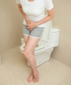
Urinary incontinence is the loss of bladder control. It is a common yet embarrassing problem. It can be as mild as releasing a small amount of urine when you laugh or sneeze, or as serious as having the urge to urinate come on so strong and fast that you don’t have time to get to a bathroom. Problems with the bladder sphincters can cause several different forms of incontinence.

One form of urge incontinence occurs when the urethra can’t hold back urine in the bladder and the bladder sphincters relax uncontrollably.

Stress urinary incontinence is another common issue. In this case, stress or damage to the sphincters or surrounding muscles can cause urinary incontinence. This is common in women after childbirth, or in men after prostate surgery or radiation therapy. Neurogenic bladder dysfunction occurs when trauma or disease of the central nervous system causes a person to lose control of their bladder sphincter muscles.

Although issues caused by malfunctioning bladder sphincters can be embarrassing, there are a variety of treatment options available. The option that works best–whether it be lifestyle changes, medication, or surgery–depends on the patient and their overall medical history. Urologists work to understand their patient’s unique needs and develop the best plan to treat their bladder problems. For more information, visit the Advanced Urology Institute website.

Ways to Pass Kidney Stones

Kidney stones can be a painful and difficult experience for the many men and women who get them. These stones are hard deposits of minerals that build up in the body and cause blockages in the urinary tract. The blockages can make it very painful and difficult to urinate. In addition to difficulty urinating, the stones can cause nausea as well as pain in the groin and abdomen. Luckily, there are many ways that urologists can help speed up the process.

Quynh-Dao Tonnu, PA-C: Physician Assistant in DeLand, FLFinding the best way to help a patient to pass a kidney stone depends on several factors. Not all patients are the same, and the size and difficulty of their kidney stones vary as well. If the stones are smaller than 5 millimeters, the urologist will want the patient to try and pass them naturally. This is the least invasive way to pass kidney stones, although it may not always be possible. Drinking lots of liquids for frequent urination is the key for this method. Urologists may also recommend movement like bicycling and jumping jacks to help dislodge the stones naturally.

If passing naturally is not an option, then medication may be the next step. Urologists will use some of the same medications that are used to treat an enlarged prostate. Medication like Flomax helps relieve pressure on the urinary tract by relaxing the muscles in the prostate. This allows for easier urine flow and can help the patient pass the stones.

Another treatment option that makes passing stones easier is called shock wave lithotripsy. This treatment uses a machine pressed up against the patient’s body that targets water waves directly at the kidney stones. With as many as 2,500 waves per treatment, these waves break the kidney stones into small, sand-like particles. The sandy remnant is then easily passed during urination.

In some cases, a more invasive treatment may be needed to remove the stones from the body. A surgeon may use a scope fitted with a medical laser to enter the patient’s urinary tract through the penis. They can then use the laser to break the stones up into smaller fragments for easier passing. This method is sure to help pass the stones, but a urologist may prefer to try more natural methods first.

There is no single method to passing kidney stones. What matters most is making the process as fast, safe and pain-free as possible. For more information about kidney stones, visit the Advance Urology Institute website.

How Do I Know If I Have a Kidney Stone

Kidney stones is the common term for the medical condition of renal lithiasis or nephrolithiasis. The stones are made up of salts and minerals that form into hard deposits inside your kidneys.

Kidney stones can be caused by many different factors and can show up in different sections of your urinary tract. They may travel from the kidneys to your bladder, and from the bladder to the ureter. Sometimes kidney stones occur when the urine is too concentrated, which allows minerals to coalesce and crystallize. Heeding early signs like painful urination and an irregular urge to pee can help you seek medical help earlier and get treatment.

Pain from Kidney Stones

How can you be sure that kidney stones are causing your pain?
Because there are other maladies that have similar symptoms to kidney stones, a visit to your urologist may save you from uneccesary grief. Some symptoms of kidney stones include:

  • Acute pain below the ribs on the side of your body or in your back. The pain may intensify or shift to various locations as the stones travel along the urinary tract.
  • Pain located in the lower abdomen or groin
  • Pain that fluctuates in severity
  • Painful urination
  • Cloudy or discolored urine (pink, red or brown)
  • Foul-smelling urine
  • Persistent need to urinate or urinating more than usual
  • Urinating in small amounts
  • Nausea and consequent vomiting
  • Chills and fever due to associated infection

You could have just one of these symptoms or several. If you are experiencing severe discomfort along with nausea or blood in your urine, you should seek medical help.

Kidney stones do not usually cause any permanent damage if treated early and correctly. You may be able to drink plentiful amounts of water, take a pain medication, and be able to pass the stone on your own. However, if the stones get lodged in the urinary tract or cause a urinary infection, more aggressive treatment may be in order.

Tests for Kidney Stones

There are several ways your doctor can test for kidney stones that will also reveal their size and precise location. These tests include:

  • Imaging tests: The imaging technology to determine if you have kidney stones includes X-rays, CT scans and ultrasounds.
    CT scans are more thorough than X-rays and result in lucid composite images of kidney stones. Ultrasound is also used to create images of the affected area.
  • Blood tests: Your doctor can determine if your have too much uric acid or calcium in the blood, which can cause kidney stones to form.
  • Urine tests: These lab tests can detect minerals that cause kidney stones in your urine. They can also reveal if you lack elements that prevent stones from forming.

Treatment for Kidney Stones

Your doctor can determine if sound-wave therapy can resolve the problem or if surgery is indicated due to stones being too large to pass, causing infection or other damage. If you are able to pass the stone on your own, saving it for your urologist to examine can help your doctor determine what causes your stones and what can be done to prevent additional ones from forming.

If you would like more information about kidney stones and their treatment, schedule a consultation at the Advanced Urology Institute location nearest you or visit the website.

What are the benefits of da Vinci Robotic Surgery According to Dr. Rolando Rivera

KEY TAKEAWAYS:

  • The da Vinci® robotic surgery technology has revolutionized the field of minimally invasive surgery, offering consistent and superior outcomes and enabling patients to recover faster.
  • The benefits of da Vinci robotic surgery for patients include reduced pain, less blood loss, reduced risk of infection or complications, less scarring, shorter hospital stays, and faster recovery.
  • The da Vinci system offers surgical professionals advantages such as improved dexterity, a wider range of motion, enhanced visualization, improved access to hard-to-reach areas, and improved outcomes that spare healthy tissue.

If you are facing surgery, you want the most effective and least invasive approach to the operation. You can rest easy when you hear that your procedure will be done with the da Vinci® robotic surgery technology.

The da Vinci approach has transformed minimally invasive surgery, enabling patients to get back to what matters sooner. Over the past 20 years, da Vinci platforms have offered consistent and superior outcomes, pioneering new capabilities in the operating room. The da Vinci robotic surgery has virtually revolutionized the field of minimally invasive surgery.

Dr. Rolando RiveraSurgical Approach with da Vinci Robotic Surgery

  1. Surgeons and operating room staff who use the da Vinci system are given comprehensive training by the manufacturer. Their professional education includes simulation for skills and procedures, along with practice and peer guidance.
  2. The da Vinci robotic surgical system offers a minimally invasive alternative to laparoscopy and also to open surgery. (Laparoscopy surgery is a procedure that uses fiber-optic instruments inserted through the abdominal wall to see and repair internal organs.)
  3. Because robotic surgery offers greater precision and control for the surgeon, and is performed by just a few tiny incisions, patients generally recover sooner.

Benefits of da Vinci Robotic Surgery for Patients

Patients will be thankful for the many benefits of robotic surgery using the da Vinci approach:

  • Reduced pain
  • Less blood loss
  • Reduced risk of infection or complications
  • Less scarring because of smaller incisions and fewer sutures
  • Briefer hospital stays/faster recovery
  • More rapid return to normal activities, including urinary continence, sexual function, and more.

Benefits of da Vinci Robotic Surgery for Surgical Professionals

To understand more about the benefits of the da Vinci system, consider these advantages. Aided by the da Vinci system’s ergonomic design, your surgeon is able to get a magnified 3-D view with high-definition while operating from a comfortably seated position. Your surgeon can operate precisely with these advantages:

  • Improved dexterity
  • Wider range of motion
  • Enhanced visualization including areas that are not visible by the naked eye
  • Improved access to otherwise almost inaccessible areas
  • Improved outcomes, sparing healthy tissue not impacted by cancer

The superior results with a da Vinci robotic surgery procedure are ideally suited for urologic conditions. At the Advanced Urology Institute, urologists such as Dr. Rolando Rivera perform minimally invasive robotic surgeries for prostate cancer, vasectomies and more complex surgeries such as reconstruction. For more information, visit the Advanced Urology Institute website.

TRANSCRIPTION: 

I’m Rolando Rivera and I’m board certified in Urology and Female Public Medicine and Reconstructed Surgery with Advanced Urology Institute.

Yes, the DaVinci machine, which is a trademark platform in general robotic surgery, is designed to improve the ability of the surgeon to get the tissues and see the structures through small incisions via laparoscopy, it’s based on laparoscopic principles.

You can get to the different organs and use 10 times magnification in a three-dimensional array, so you can see things very close and very magnified. And the robotic platform allows you to have more precision, takes away the surgeon’s tremor because the instruments do all the movement that you guide in a console, so it allows you to do more refined treatments and surgical techniques. Well, because you’re not opening big incisions and you’re creating less trauma, less trauma leads to less inflammation, then pain is less of an issue. And whenever you have small incisions, less trauma, less inflammation, you’ll have quicker recovery.

REFERENCES: 

What Causes Kidney Stones

Kidneys perform the function of filtering blood to remove waste from it. This waste is mostly water that contains salts such as sodium, calcium and potassium, all collectively referred to as urine. Kidney stones form when these salts are so concentrated in the urine that they crystallize into clumps, that can be as small as a grain of sand or larger.

Ordinarily a kidney stone causes no symptoms until it begins to move in the kidney or to other parts of the urinary system. A small kidney stone can be passed painlessly by urination. Bigger kidney stones, however, can be very painful to pass. But kidney stones are treatable and treatment should help you avoid the painful experience.

Causes of Kidney Stones

Kidney stones result from the clumping together of salts. Most kidney stones are calcium oxalate stones that form from calcium salts and oxalates. Oxalates occur naturally in some fruits, vegetables and nuts. There are also uric acid stones, which form due to lack of fluid in the body or a significant loss of fluid. Risk factors for the formation of kidney stones include:

1. Lack of Water/ Dehydration
This is without a doubt the most common cause of kidney stones. Stones form when urine salts occur in very high amounts in urine. Drinking adequate amounts of water can dilute the urine and reduce the concentration of salts. People in hot areas should take special care to drink a lot of water because they lose a lot of it in perspiration.

Dr. Rolando Rivera 2. High sodium diet
Many kidney stones have an element of calcium. Too much sodium increases the amount of calcium that kidneys have to process, thereby making it more likely for stones to form. Animal proteins such as red meat and shellfish, also have a reputation for making urine acidic, thereby increasing the possibility of having uric acid stones.

3. Personal or family History
A person who comes from a family with a history of having kidney stones is at an increased risk of having them. Similarly, a person who has had kidney stones before is very likely to suffer a recurrence.

4. Digestive illnesses and surgery
Serious inflammations of the digestive system, chronic diarrhea and gastric bypass surgery all can negatively affect the absorption of fluids and calcium by the body. This makes it more likely for the patient to have kidney stones.

Kidney stones are very common despite the fact that they can be prevented by staying hydrated and watching your diet. If you have them, you should consult a doctor immediately so treatment can be administered promptly. The Advanced Urology Institute offers effective treatment for kidney stones. Its staff of specialists are available to help with diagnosis and treatment and can answer any questions about prevention of kidney stones. For more information, visit the Advanced Urology Institute website.

Causes of a Weak Urine Flow

The inability to start or maintain urine flow, causing dribbling or weak urine flow, is called urinary hesitancy. It can occur at any age for all genders, but it is by far most common among aging men. Urinary hesitancy develops gradually but if left untreated, it can lead to a complete inability to pass urine, also known as urine retention. Due to its prevalence among older men, weak urine flow has always been associated with an enlarged prostate gland, a condition very common in older men. This, however, is not the only cause of a weak urine flow.

Causes of a weak Urine Flow

1. Benign Prostate Hyperplasia (BPH)

Benign prostate hyperplasia is the medical term for an enlarged prostate. As a man ages, the prostate grows to a size larger than what is normal. The prostate’s location around the tip of the urethra means that the enlarged prostate presses on the urethra and blocks the passage of urine. This whole or partial obstruction slows down the flow of urine. BPH is the main cause of a weak urine flow in men over the age of 45.

2. Underactive Bladder (UAB)

Dr. Jonathan Jay: Naples, FLAn underactive bladder is a medical condition characterized by weak urine flow and an inability to empty the bladder completely. The condition is best understood as the opposite of the overactive bladder (OAB) which has received wider attention and is known to cause urinary incontinence, the urgent and frequent urges to urinate. Patients suffering from an underactive bladder have a diminished sense of when their bladder is full and are not able to contract the bladder fully. The condition can occur when there is damage to the bladder peripheral pathways or to the lumbosacral spinal cord. It is also common in patients suffering from diabetes mellitus, neurological diseases, Parkinson’s disease and pelvic fractures.

3. Bladder Outlet Obstruction (BOO)

As the name suggests, this is a condition that occurs when there is a blockage at the neck or the base of the bladder. The blockage completely prevents or reduces urine flow from the bladder. It occurs in both women and men, but is more common in older men. It can be caused by bladder stones, scar tissue in the urethra, bladder cancer or an enlarged prostate.

It is clear that weak urine flow has a variety of causes. It is very important to see a urologist to determine the exact cause of a weak flow. An appropriate plan of treatment can be set up once it has been determined what is creating the problem. It is recommended that you consider seeking out trained, experienced and board approved urologists if you are having problems with weak urine flow.

At AUI, we offer patient-friendly and multidisciplinary urology services for a wide range of problems, including weak urine flow. For more information, visit the “Advanced Urology Institute” site.

For more information, visit the “Advanced Urology Institute” website.

Prostate Problem Warning Signs


At Advanced Urology Institute, we frequently see first-time visitors with symptoms of advanced stage prostate problems. For us, that is quite heartbreaking because it means the patients come too late, when only limited treatment options are available for their conditions.

As urologists, we always want the best for our patients. We want to see them leave when they can pee better and are free from the embarrassment of accidental urine leaks. And because early detection and treatment of prostate problems — whether prostate cancer or non-cancerous condition — improve the chances of cure and of long-term survival, we always encourage men to be more mindful of their bodies, especially when it comes to their urinary function and habits. By doing that, they are able to detect warning signs of prostate issues early and can seek treatment.

Warnings signs of prostate problems include:

  1. Frequent urination or frequent urge to pass urine.
  2. Passing urine more often than usual, particularly at night.
  3. Pain, discomfort or burning sensation when passing urine.
  4. Dribbling urine
  5. Weak or interrupted urine streams.
  6. Accidental urine leakage.
  7. Blood in urine or semen
  8. Frequent stiffness or pain in your lower back, rectal area, hips, upper thighs or pelvic area.
  9. Difficulty or inability to urinate
  10. Trouble with starting or stopping your urine stream.
  11. Painful ejaculation
  12. A feeling that you aren’t able to empty your bladder completely.
  13. Swelling of lower extremities.
  14. Paralysis or weakness in lower limbs.
  15. Inability to pass urine while standing up.
  16. Loss of appetite and weight, fatigue, nausea and vomiting.

It is important to see a urologist immediately if:

  1. You find urination difficult, abnormal or painful. The doctor will examine your prostate gland to find out if it is inflamed, enlarged or has a cancer.
  2. You have frequent urination, urinary retention, blood in urine, dribbling or slow flow of urine, problems starting a urine stream, or repeatedly urinate urgently.
  3. You have a chronic pain in your pelvic, lower back, upper thigh or other areas of your lower extremities. While any unexplained ongoing pain in these areas may have various causes and always merits medical attention, seeing a urologist may help detect whether or not you have prostate cancer.
  4. You have swollen legs, weakness in your legs or trouble walking.
  5. You have unexplained weight loss.

As urologists, we have several options for tackling prostate problems, but our interventions normally depend on the severity of the symptoms, type of condition and how it is impacting your overall quality of life. For example, if the prostate condition is not severely affecting your quality of life and you have no complications (such as bleeding, bladder stones or urinary infections), the decision to treat the problem is often optional and left for you to make.

That means if you aren’t bothered enough to undergo a procedure or take medicine for the condition, then you’ll only need frequent follow-up with your urologist to check whether your symptoms remain stable over time and your bladder continues to empty well. But if you already have complications or your bladder is holding increased quantities of residual urine after urination, then we often begin treatment immediately.

To help you pass urine better, we may offer medications such as alpha blockers, 5-alpha reductase inhibitors or a combination of drugs. But minimally invasive surgical procedures such as TUMT (transurethral microwave thermotherapy) and TUNA (transurethral needle ablation), water-induced thermotherapy, PVP (photoselective vaporization of prostate), and HoLAP (holmium laser ablation of prostate) also may be considered. For more information on diagnosis and treatment of prostate problems, visit the “Advanced Urology Institute” site.

How are Kidney Stones Treated?

Kidney stones are a common cause of agonizing and debilitating pain in men and women. In the United States, the stones account for over one million hospital visits and more than 300,000 emergency room visits every year. When patients present with kidney stones, the treatment administered usually depends on the type, size and location of the stone and on the severity of symptoms. Apart from administering treatment, the urologist investigates the underlying cause of the stones and recommends ways of preventing a recurrence.

Spontaneous passage

Kidney stones smaller than 4 mm in diameter are often passed on their own in urine and may be treated at home. While such stones may be painful, the pain often lasts only a few days and usually disappears soon after the stone is passed. So, depending on how bad your symptoms are and how long you’ve had the symptoms, you may not be given any form of treatment and just wait for stones to pass in urine. It usually take up to six weeks to do so.

However, you should only do this if the pain is bearable, there is no sign of infection or kidney blockage and the stone is of a size that can pass on its own. As you wait for it to pass, you’ll need to drink plenty of water and take pain medication to help you manage the discomfort. If you suspect that you have a kidney stone, speak with your doctor to see if you need immediate treatment or if you can wait for it to pass spontaneously.

Medications

There are a number of medications that increase the chance of passing kidney stones. For instance, tamsulosin is commonly given to people with kidney stones to help relax the ureter and make it easier for stones to pass. Apart from medications to boost stone passage, your urologist may prescribe anti-emetic (anti-nausea) medication to reduce nausea and vomiting as you wait for the stone to pass. And if you are in severe pain, your doctor may give you 1-2 pain injections and then prescribe some painkillers and anti-emetics for you to take from home.

Surgical procedures

If the pain is so much that you can’t wait for the stone to pass in the urine, you’ll require a surgical procedure to remove it. Surgery is also necessary if the stone is too big to pass on its own or is hampering kidney function. Kidney stones may be removed surgically if they are causing repeated urinary tract infections or are blocking the normal flow of urine.

Surgical procedures to remove kidney stones include extracorporeal shock-wave lithotripsy (ESWL), ureteroscopy and percutaneous nephrolithotomy (PCNL). These procedures are usually chosen by urologists depending on the size, type and location of the stones.

1. Extracorporeal Shock-Wave Lithotripsy (ESWL)

This procedure is the most frequent way of treating stones that can’t pass spontaneously in urine. High-frequency waves (X-rays or ultrasound) are directed at the stone to break it into smaller pieces that can pass in urine. Often the tiny pieces require a few weeks to pass out in urine. While ESWL is 99 percent effective for kidney stones up to 20 mm in diameter, more than one session is usually necessary for the treatment to be successful.

2. Ureteroscopy

For kidney stones that are lodged somewhere in the kidney or ureter, ureteroscopy (also called retrograde intrarenal surgery) may be necessary. The procedure involves directing a long, thin telescope (called ureteroscope) through the urethra, into the bladder, then into the ureter or kidney where the stone is located. If the stone is stuck in the kidney or upper ureter, the urologist uses flexible telescopes for this procedure, but rigid telescopes are ideal for stones stuck in the lower parts of the ureter.

The ureteroscope helps the urologist to reach the stone without making an incision. After reaching the stone, the doctor either can use another instrument to remove it or direct laser energy on it to break it into smaller pieces that can pass naturally in urine. A stent (plastic tube) may be inserted temporarily into the bladder to drain out the stone fragments.

3. Percutaneous nephrolithotomy (PCNL)

For kidney stones that are too large (21-30 mm in diameter), percutaneous lithotripsy is the treatment of choice. During the procedure, a half-inch incision is made in the side or back, just big enough to allow passage of a telescopic instrument (called nephroscope) into the area of the kidney where the stone is located. The nephroscope is used either to pull out the stone or break it up with pneumatic energy (or laser) and suction out the pieces. In fact, it’s the ability to suction out tiny stone pieces that makes this procedure ideal for larger stones.

Kidney stones also can be removed through open surgery, laparoscopic surgery or robotic surgery. But this is only done when the less-invasive procedures fail. Routine surgical procedures for kidney stones require shorter recovery period and you can usually return home the same day after the procedure and resume normal activities in 2-3 days. If the urologist inserts a stent after a procedure, it is removed 4-10 days later. During treatment, you also may be provided with a strainer that you can use to collect stone pieces that pass in urine for laboratory testing and to enable the urologist to recommend appropriate ways of preventing stone recurrence.

At Advanced Urology Institute, we offer shockwave lithotripsy, ureteroscopy and percutaneous nephrolithotomy routinely, and perform robotic and laparoscopic procedures for kidney stones when necessary. We perform blood tests and 24-hour urine analyses for every patient to identify the cause of kidney stones in order to provide the right treatment. We also design prevention strategies tailored to each patient, including personalized dietary recommendations based on results of 24-hour urine analysis. Our aim is to always ensure that our patients properly understand why they have kidney stones and make the necessary lifestyle changes to prevent a recurrence. For more information on kidney stones and how they are managed, visit the “Advanced Urology Institute” site.

What Does the Prostate Gland Do?

The prostate, found only in men, is a tiny, walnut-sized muscular gland in front of the rectum and just below the bladder. It surrounds the urethra, which transports urine to the penis from the bladder, and is essential for the normal functioning of the male genitourinary system. The prostate makes prostatic fluid, which forms a substantial portion of semen. During ejaculation the prostate contracts and closes off the opening between the urethra and bladder, ensuring that prostatic fluid squirts into the urethra and semen is pushed out at speed.

So what does the prostate do?

The prostate is not an essential organ for life, but it’s quite crucial for reproduction. It plays an active part in reproduction by secreting the prostatic fluid, which forms part of healthy semen — the perfect environment for the transit and survival of sperm. Healthy semen includes the enzyme PSA (often measured during screening for prostate cancer), together with other substances secreted in the prostate and seminal vesicles, such as citrate, zinc and fructose, which supply sperm with the energy to travel to the egg. There are also various antibodies in semen to protect sperm and the urinary tract from bacteria and different pathogens.

What are the constituents of prostatic fluid?

The prostate secretes a milky fluid, the prostatic fluid, which makes up about 30 percent of the total fluid that’s ejaculated. The prostatic fluid contains various ingredients, such as enzymes, citric acid and zinc, which keeps sperm alive and protects them and the genetic codes they carry. PSA (prostate-specific antigen) is one of the enzymes in the fluid that, after ejaculation, makes semen runnier and helps sperm travel through semen more easily, which increases the likelihood of successful fertilization of an egg. While prostatic fluid is weakly acidic, the other constituents of semen turn it alkaline overall, which helps to counteract vaginal acidity and prevent damage of sperm.

Growth of the prostate

The prostate grows bigger as men age. Typically the prostate undergoes growth during adolescence driven by the male hormone, testosterone, and the hormone’s byproduct, dihydrotestosterone (DHT). Testosterone hormone is primarily produced in the testes, but smaller quantities also can be secreted in the adrenal glands found just above the kidneys. By the age of 40 the prostate might have grown from a walnut-sized to apricot-sized gland. And by the age of 60 it might have reached the size of a lemon.

Benign prostatic enlargement (BPH)

While the prostate is strategically located to deliver the prostatic fluid and squeeze things along during ejaculation, its position around the urethra makes it a liability when it grows or gets bigger. A swollen prostate will compress the urethra and irritate the walls of the bladder, resulting in interference with normal urine flow. In fact, over 50 percent of all men in their 60s have symptoms of prostate enlargement, a condition called benign prostatic hyperplasia (BPH). And by the age of 70 to 80, a man’s risk of BPH increases to 90 percent, with symptoms such as leaking or dribbling urine, frequent urination, weak or stuttered urine stream.

While the size of your prostate will not always influence how severe the obstruction or symptoms are, BPH can cause serious complications over time, such as urinary tract infections, bladder damage, kidney damage, incontinence and bladder stones, due to bladder strain and urinary retention. So it’s important to see a urologist as soon as you have urinary issues so BPH can be detected early and treated.

Prostate cancer

Another health problem associated with the prostate is prostate cancer, which is the most frequent cancer in men. It occurs in 1 out of 7 men over their lifetime, and more than 200,000 men are diagnosed with the cancer annually. Prostate cancer is frequent in men age 50 and older, but the largest number of cases is found in men 70 to 80 years old. About 3.8 percent of men diagnosed with the cancer die of the disease, while 7 in 10 newly diagnosed patients currently survive past 5 years. As doctors, our role is to screen for the cancer, detect aggressive forms early and prevent any problems that may occur due to obstruction.

So what’s your role?

Tell your doctor about your urinary problems as soon as possible. The symptoms may not only suggest BPH, but also may indicate a more serious condition. With a prompt visit to the doctor, a more serious condition such as prostate cancer can be ruled out.

Want to know more about BPH, prostate cancer and other urological problems? Visit the “Advanced Urology Institute” site.