Women may feel apprehensive about visiting a urologist due to its personal nature. Learn more from Dr. Szell, urologist in Palm Harbor, FL.
Continue readingKidney Stones: Risk Factors and Preventions
 The prevalence of kidney stones in the United States has increased over the last decade. As many as 1 in 10 Americans have a kidney stone at some point in their lives, and every year more than half a million Americans go to emergency rooms for kidney stone related complications.
What are kidney stones?
A kidney stone is a small, hard deposit that forms in the kidneys. Stones occur when the urine concentration of crystal-forming substances—such as calcium, oxalate and uric acid—is more than the fluid in the urine can dilute.
They begin as small crystals and grow into larger masses (stones), which then make their way through the urinary tract. Unfortunately, a stone can get stuck on its way out of the urinary system, resulting in an unbearable pain that comes in waves until the stone eventually passes.
What causes kidney stones?
Genetics is one of the risk factors. If you have family members who had kidney stones, you are at a higher risk of having them yourself. Your risk is also higher if you have had kidney stones in the past.
Dehydration is another major cause of kidney stones, which is why more kidney stones occur in the summer. In fact, kidney stone frequency is known to vary by geographic location, with warmer climates having the highest rates of stone formation.
What you eat and drink makes a huge difference. Drinking enough fluids to make over two liters of urine a day reduces the risk of stone formation. Actually, as a rule, you should always check your urine for signs of dehydration. If your urine is dark or yellow, then you are not drinking enough fluid and run the risk of having stones.
Factors that increase dehydration will contribute to kidney stone formation. For instance, excess salt or sodium in food, such as in processed or fast foods, increase dehydration as the excess salt requires a lot of fluid to excrete. So reducing the sodium in your diet will minimize your risk of stone formation.

How do you know that you have kidney stones?
Kidney stones cause pain by getting stuck in the urinary system. Since the kidneys continue to make urine, which in turn can’t get out due to the blockage, the urine builds up, stretches the kidneys and leads to severe pain.
You will know you have kidney stones when you have severe, excruciating pain that comes in waves. The pain typically occurs in the back and does not get better with a change in position. Patients who have had kidney stones and also delivered children report that the stones are more painful than giving birth. In addition to pain, you may have fever, nausea, and even vomiting.
Kidney stones may require a trip to the emergency room if you have severe pain, nausea, vomiting, and a fever greater than 100.3 degrees. These symptoms constitute a urological emergency because they signal both a blockage and an infection. With the blockage preventing antibiotics from getting out via urine, you can get very sick, very quickly; hence the need for emergency care.
Emergency treatment with IV fluids at a hospital may be necessary if you are having nausea and vomiting to the point of dehydration. Emergency care is also appropriate when you have pain that cannot be alleviated by over-the-counter pain medicine.
What is the treatment for kidney stones?
The treatment for kidney stones depends on the size and location of the stone, and on the clinical stability of the patient. The most common approach is medical expulsion therapy—a conservative approach for healthy patients with stones that are small enough to pass on their own and with no fever or other signs of infection.
With medical expulsion, you are encouraged to drink a lot of fluid to help the stone pass on its own. You are also given medications to control the pain and to accelerate passage. If the stone is 5 millimeters or smaller (about half of your thumbnail), there is a 50% chance it will pass on its own and you will avoid surgery.
If you have severe pain, fever, chills and an inability to drink fluids, you may not qualify for medical expulsion therapy. In that case, a surgical procedure may be needed. There are two common surgical options: (1) ureteroscopy or laser lithotripsy, and (2) extracorporeal shockwave lithotripsy.
Ureteroscopy and laser lithotripsy are fancy ways of saying you go to sleep, a camera is inserted through your urethra to the stone, and a laser is used to break the stone into smaller fragments for removal. Extracorporeal shockwave lithotripsy means you go to sleep and sound waves are sent through your skin to fracture the stone into small pieces that can pass on their own in urine.
The advantage of shockwave lithotripsy is that nothing goes into your body, making it less invasive. However, the disadvantage is that the stone fragments still have to pass on their own, a process which can be painful and uncomfortable.
How do you prevent kidney stones?
1. Increase calcium intake
There is a misconception that increasing dietary calcium increases the risk of calcium oxalate stones. This is not true. In fact, eating more calcium rich foods, such as milk or cheese, ensures the oxalate in the diet binds to calcium. When oxalate binds to calcium in the intestines, it is not absorbed in the bloodstream and ends up in stool.
2. Reduce oxalate rich foods
Foods high in oxalate, such as beets, chocolate, tea, coffee, spinach, kale, rhubarb, nuts and beer contribute to stone formation. You may have to eat smaller portions of these foods alongside calcium-rich foods or avoid them altogether.
3. Stay hydrated
Drinking plenty of water will ensure that substances in your urine are diluted and cannot form crystals. As a rule, strive to drink enough water to pass two liters of urine every day—which is drinking roughly eight standard 8-ounce cups per day. It also helps to include some citrus beverages, such as orange juice and lemonade, as the citrate in these beverages helps to block stone formation.
4. Reduce sodium intake
A high salt or sodium diet increases the amount of calcium in your urine and triggers stone formation. Excess salt also wastes the fluid you take as a lot more fluid is necessary for salt-water balance. Make sure to limit your daily sodium intake to 2300mg or less to reduce your risk of kidney stones.
5. Minimize intake of animal protein
Animal protein, such as red meat, eggs, poultry, or seafood, increases the level of uric acid in the body and may cause kidney stones. A high protein diet will also reduce your urinary citrate—the chemical in urine that prevents stones from forming. You can limit animal proteins or replace them with plant-based proteins.
At Advanced Urology Institute, we offer a range of treatments for kidney stones depending on the severity of symptoms and the type, size and location of the stones. We also run tests to find out why they form and give you advice on how to prevent them.
If you or a relative has had kidney stones, consider meeting with one of our urologists for specific ways to reduce your risk. For more information on kidney stone causes, risk factors, diagnosis, treatment and prevention, visit the Advanced Urology Institute website.
Listen to the Podcast to learn more about Kidney Stones, Click here
Advantages of da Vinci Surgical System
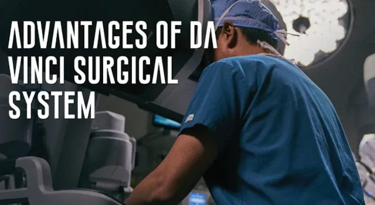
The da Vinci Surgical System is a minimally invasive procedure where the operation is performed with the help of robotic instruments inserted through minor incisions. The surgeon sits at a console that offers a three dimensional view of the patient’s anatomy and full control over the surgical instruments.
The da Vinci system provides a suitable alternative to the more traditional open surgery where a surgeon has to make large incisions through skin and muscle to reach the affected organ. It also is an improvement to other minimally invasive procedures, such as laparoscopy. Now a patient has the opportunity to choose robot assisted laparoscopy over conventional laparoscopy.
Advantages of the da Vinci Surgical System
1. Increased precision
The precision with which miniaturized robotic instruments move through the body is comparable to that of a scalpel in a surgeon’s hand. Robotic instruments can access parts of the anatomy that are delicate or hard to reach with no risk of accidental puncture or laceration. The margin of error is very low because precision increases accuracy.
2. Reduced blood loss, pain and scarring
Blood loss, pain and scarring are incidental to any surgical procedure. Blood loss is especially critical and it is a significant contributor to mortality rates in procedures such as hepatic and thoracic surgery. The da Vinci Surgical System requires very little cutting and the incisions made are very small. Advantages of the smaller incisions include less pain, smaller amount of blood loss and a shorter healing period after the surgery. The high definition cameras and the 3D view in the da Vinci Surgical System also offer increased visibility, which reduces the chances of puncturing blood vessels or any other part during the surgery.
3. Faster recovery
Patients who undergo surgery using the da Vinci system report faster recovery. They require less after care and the risk of post surgery complications is remarkably reduced. This is especially the case with post surgery infections, which are a concern in cases of open surgery because of the larger wounds. Patients undergoing this kind of surgery realize the objectives of their treatment quickly and they can return to their normal activities with no problems.
Conclusion
The da Vinci Surgery System is available for almost all forms of surgery, including gynecological, kidney and colorectal. Areas such as urology are known to have been at the forefront of adopting this system to urological surgeries and to date, most urologists, including the members of the Advanced Urology Institute, champion the use of the da Vinci Surgery System.
For more information about the da Vinci Surgery System, visit the “Advanced Urology Institute” website.
2 Effective Treatment Options for Prostate Cancer
Prostate cancer can be treated and managed in a number of ways. While the preference of the patient is given priority, it should be tempered with the advice of a trained urologist. A urologist can offer advice on what method is appropriate depending on the age of the patient, the patient’s family history and the natural progression of the disease. The patient also needs to be fully advised of the side effects of any form of treatment before agreeing to undergo the treatment. For any given case of prostate cancer, there are always at least two treatment options available.
1. Surgery
A patient with prostate cancer can choose to have the prostate surgically removed to clear the cancer from the body. The procedure is known as Radical Prostatectomy. It is most appropriate in cases where the cancer is localized and has not spread beyond the prostate. However, even when the cancer is localized, a urologist will determine the progression of the disease before recommending surgery. Low risk localized prostate cancer is unlikely to progress and a radical prostatectomy is unnecessary. On the other hand, when the cancer is aggressive and is likely to result in death if untreated, surgery is definitely the most appropriate choice.
Radical prostatectomy is recommended for patients under the age of 75 , or those with a life expectancy of at least ten years. This is because they are more likely to preserve their sexual and urinary functions after the surgery and they have a stronger chance of outliving any side effects the surgery might have.
2. Radiation Therapy
Radiation therapy, or radiotherapy is the use of radiation in high doses to kill malignant cancer cells or slow their development. Unlike radical prostatectomy, it can be used to treat localized prostate cancer and even advanced prostate cancer. It may be applied in combination with other treatment options such as hormone therapy. It may even be applied if a patient undergoes a radical prostatectomy but the procedure fails to eliminate the cancer fully or if the cancer recurs.
Radiation therapy can be administered externally or internally. When done externally, it is referred to as external beam radiation and is very much like having an X-ray. When administered internally, it is referred to as brachytherapy or internal radiation therapy. In this procedure, a radiation implant is placed inside the body near the affected organ. After a while, the implant ceases to produce radiation. The implant, however, remains in the body.
Both radical prostatectomy and radiation therapy are suitable treatment options and choosing between them may seem a little daunting. The professional opinion of a urologist can help by pointing out the finer points of each choice. The patient also may research the subject by reading up on the various options. There are many sites that offer reliable material on this subject.
For more information about treatment options for prostate cancer, visit the “Advanced Urology Institute” website.
Robotic Surgery Effective in Partial Nephrectomy
Robotic partial nephrectomy involves using an advanced surgical robot to remove part of the kidney, usually the portion with a tumor. Initially, robotic surgery enjoyed tremendous success with surgical removal of the prostate (prostatectomy), but in recent years its usage in kidney operations also has yielded remarkable results. In fact, robotic partial nephrectomy has become the preferred treatment option for most patients with benign kidney tumors, small renal masses and early-stage cancer. During the procedure, tumors are removed with the least possible disruption of the rest of the kidney — a nephron-sparing approach that maximizes post-operative kidney function.
Why is the da Vinci surgical system suited for partial nephrectomy?
The da Vinci surgical robot provides superior maneuverability that is suited for the delicate slicing, cutting and stitching involved in the removal of a portion of the kidney. The surgical robot offers a three-dimensional view of the targeted area, allowing for a broader range of motion of the surgical devices. Urologists using the robot find it much easier to make the complex maneuvers required during the procedure.
Since it uses smaller incisions and doesn’t involve making cuts through bone or muscle, the da Vinci partial nephrectomy causes less scarring and minimal trauma to patients. The recovery time is typically only 2 weeks compared to the 6-8 weeks recovery time after open kidney surgery. Likewise, blood supply to the kidney is blocked for a shorter duration, leading to less overall blood loss and quicker recovery compared to laparoscopy.
How is the robotic partial nephrectomy performed?
During robotic partial nephrectomy, the surgeon makes a series of tiny incisions in your abdomen. The camera and robotic surgical instruments are inserted through these incisions. To create enough room for manipulation of the surgical instruments and enable easy access to the cancerous tissue, the abdomen is inflated with gas (carbon dioxide gas). The doctor then moves the colon away from the kidney and trims off the fat covering the kidney to expose the kidney surface.
With the kidney exposed, the surgeon halts the blood flow to the kidney temporarily to prevent potential bleeding as the tumor is cut and the remaining portion of the kidney sutured together. At the end of the procedure, the urologist reconstructs the kidney, restores blood flow and then inspects the kidney carefully to make sure there is no bleeding.
Who should undergo robotic partial nephrectomy?
The da Vinci partial nephrectomy is the surgical treatment of choice for patients with smaller kidney tumors, usually not bigger than 4 cm in size. However, even patients with tumors ranging from 4 cm-7 cm also may undergo the procedure if they are to be treated in certain areas. Similarly, robotic partial nephrectomy is appropriate in cases where removing the whole kidney could trigger kidney failure or the need for dialysis.
At Advanced Urology Institute, we perform hundreds of robotic partial nephrectomy every year with amazing results for our patients. The procedure takes a short time, reduces the problems caused by benign or small kidney tumors and is effective in helping patients recover from kidney cancer. The minimally-invasive nature of the procedure guarantees less scarring, minimal trauma and quicker recovery for our patients. But we always ensure that patients are closely monitored for post-operative pain and complications, accomplishing cancer-free and happier lives for our patients. For more information on treatment of kidney cancer and other urological problems, visit our “Advanced Urology Institute” site.
What is MRI with Transrectal Ultrasound Fusion-Guided Prostate Biopsy
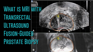
 Prostate cancer has a new standard of care in MRI-guided fusion biopsy with transrectal ultrasound. While a prostate biopsy has been the only way to get a definitive diagnosis of prostate cancer, it has only been working if cancer cells are identified in the sample tissue. But in some cases, such as when the tumor occurs at the top surface of the prostate or other unusual locations, a biopsy may not give a correct diagnosis. For instance, the standard TRUS (transrectal ultrasound) guided biopsy in which tissue samples are collected from the prostate in a systematic pattern gives a negative result with tumors located in unusual areas of the prostate. About 15-20 percent of tumor locations can be missed by the biopsy needle.
What makes the MRI-ultrasound fusion biopsy more definitive?
The MRI-ultrasound fusion approach is an improvement on the traditional 12-core TRUS, which involved taking biopsies from twelve prostate areas where the cancer is considered more likely to occur. With the TRUS biopsy, about 70 percent of men who have a negative biopsy result are not essentially free of the cancer. The MRI-ultrasound fusion technology blends the superior imaging capability of the high-definition multi-parametric (mp) MRI with real-time ultrasound imaging. There is better visualization of the suspicious areas of the prostate where the cancer may occur that may not be visible on ultrasound alone. The fusion-guided biopsy detects almost twice as many prostate cancers in all stages as the standard TRUS biopsy.
The ability of MRI-ultrasound fusion-guided biopsy to create a three-dimensional (3D) map of the prostate ensures that doctors are able to see the targeted areas of the prostate better and perform more precise biopsies. The technology uses a machine known as UroNav developed by Invivo, which is supplied with sophisticated software to produce super-detailed MRI images and fuse them with the ultrasound images generated by a transrectal probe administered on the patient in an outpatient setting. The resulting images enable the examining physician to direct biopsy needles with pinpoint accuracy and to easily access any lesions or suspicious areas revealed by MRI. The technology allows the urologist to hit the target spot more accurately and improves cancer detection rate. In fact, it is primarily used for men who have an ongoing suspicion of prostate cancer, such as those with consistently elevated PSA, but whose TRUS biopsy results are repeatedly negative.
Fewer biopsies, more accurate detection
The fusion-guided biopsy is a very targeted approach in which biopsies are performed only in highly suspicious areas of the prostate appearing in the MRI image. As a result, significantly fewer biopsies are done with the MRI-ultrasound fusion than with the traditional TRUS technique, minimizing the adverse effects that often accompany repeat biopsies. Multiple prostate biopsies can lead to complications such as bleeding, infection, urinary retention problems, sepsis or even death.
In spite of fewer biopsies, the MRI fusion approach increases the rate of detection of aggressive prostate cancer. The extensive MRI images obtained before the biopsy helps highlight both high-risk and intermediate-risk cancers often missed by traditional TRUS biopsy. With MRI-ultrasound fusion, the likelihood of detecting cancer increases as the grade of the tumor increases. The use of MRI fusion biopsy helps to avoid metastatic disease by finding cancer before it spreads to other areas of the body.
Improved cancer differentiation
Through MRI fusion, doctors are able to more accurately differentiate cancers that require treatment from the ones that should undergo watchful waiting (active surveillance). Fusion technology is able to show higher-risk cancers and does not highlight the insignificant low-grade tumors, making it less likely for urologic oncologists to over-treat indolent and low-grade cancers. A number of prostate cancers are low-grade, non-aggressive and do not cause problems at all and treating them through chemotherapy, radiotherapy or surgery can impair the quality of life or even cause death. MRI fusion effectively saves patients from the adverse effects of treating low-grade tumors. Fusion technology eliminates up to 50 percent of prostate cancer treatments that are unnecessarily administered on low-grade cancers.
At Advanced Urology Institute, we have adopted the MRI-ultrasound fusion biopsy and changed the way we screen, evaluate and diagnose prostate cancer. It has become our standard for detecting prostate cancer and we believe in the next few years it will be the gold standard for detecting the cancer. We are proud that it offers a higher detection rate, superior accuracy and reduces the rate of repeat biopsies — making our practice one of the best places for detection and monitoring of the cancer. It helps us deliver the best treatment outcomes for our patients.
If you think you are at high risk of prostate cancer or already have started experiencing some symptoms, let us show you how the precision of our high-definition MRI fusion machine, the expertise of our skilled physicians in MRI fusion biopsy and the know-how of our radiologists proficient in multi-parametric MRI imaging can help you. For more information on the treatment and diagnosis of prostate cancer, visit the “Advanced Urology Institute” site.
What Does Dr. Nicole Szell Say About Women’s Urinary Incontinence?
Talking With Your Doctor About Erectile Dysfunction
Do you struggle to have an erection when you want it most? Does your penis fail to get stiff enough for satisfactory sexual intercourse? Or does it get rigid only for a short while and then lose rigidity leaving you humiliated and apprehensive? You don’t need to worry too much if you have experienced this only occasionally. After all, almost every man has failed to achieve or maintain a good, rigid erection at some point in his life, particularly when stressed, tired, anxious or drunk. But if you have had frequent, frustrating and distressing failures to have or maintain an erection, then you may have a problem and need some sensible and sympathetic advice from a urologist.
Why Talk To a Urologist
Erectile dysfunction is a tough experience, but discussing it is even tougher and more embarrassing. You are likely to feel hesitant or scared to talk about ED with a doctor. But even with all the awkwardness you may feel in trying to open up and speak honestly about your erection troubles, talking to a urologist is probably the best step you can possibly make. A urologist is used to dealing with ED, perhaps attending to more than a half dozen cases every week. Your case would be just a routine matter to the urologist. More importantly, your erectile dysfunction may be the first sign of a very serious medical problem. A timely chat with a urologist can enable early diagnosis and treatment of a life-threatening condition.
Honest Useful Conversation
When you visit a urologist, you will meet a listening and caring professional dedicated to helping men have normal sex lives. The doctor will help you to explore the symptoms from when you first noticed changes in your erections to any medications you might be taking that could be causing your problem, your lifestyle habits and the severity of your symptoms. During this chat, being brutally honest and open with the urologist will ensure that the cause of your problem is figured out quickly and accurately. The physician also will conduct a physical exam focused on the health of your heart, nervous system, blood vessels and genitals, followed by appropriate urine and blood tests. The doctor then will be in a position to know the cause of your erectile dysfunction and to recommend the right treatment for you.
Exploring Treatment Options
Urologists are skilled in treating underlying medical conditions such as high blood pressure, atherosclerosis, heart disease, chronic kidney disease, diabetes, Peyronie’s disease or multiple sclerosis. In most cases, erectile dysfunction quickly resolves once the underlying cause is treated. Your urologist also may suggest lifestyle modifications such as reducing alcohol intake, losing some weight, stopping cigarette smoking and increasing physical activity to boost your recovery. But if these interventions do not work, the urologist may explore other treatment options, such as oral medications (like tadalafil, vardenafil or sildenafil), injectable medicines (like alprostadil), and vacuum pumps, and penile implants (bendable or inflatable). As a last resort, surgical repair (penile revascularization) of the veins or arteries of the penis may be considered.
No Longer a Secret
In the past, men didn’t have open and frank discussions about their erectile problems because sexual intercourse was often shrouded in mystery and secrecy. But with the availability of effective treatments for ED, that now seems imprudent. With the availability of innovative and highly effective treatments for erectile dysfunction, it is important to talk to your doctor about your problem and to know your options.
Are you having problems getting or maintaining an erection? Why not make a bold decision today to see a urologist? At Advanced Urology Institute, we offer cordial and compassionate consultations for men having erectile problems. Our skilled and experienced urologists will listen to you, do a thorough physical examination, run appropriate tests and diagnose your condition accurately. We have the necessary diagnostic tools to determine any kind of underlying medical conditions that may be causing your erectile dysfunction. Make an appointment now at AUI, have a candid chat with a urologist and have your problem fixed. For more information on the treatment of erectile dysfunction, visit the “Advanced Urology Institute” site.
Webb McCanse Becoming a Urologist
Becoming a Urologist
The opportunity to work in the Navy was very attractive to me. So I pursued medicine as a path to serving my country. With the United States
Navy taking care of my fees, I completed my medical school training at the University of Kansas and joined the University of Nebraska’s Medical Center for a six-year urology residency. Upon completion, I served in the United States Naval Hospitals of Pensacola and Guam, with sporadic assignments in Cuba and Okinawa, Japan. Following a satisfying naval service, I moved to Advanced Urology Institute.
Areas of Expertise
My extensive training exposed me to a number of advanced technologies and medical procedures. I am an expert in minimally invasive surgical procedures, particularly laparoscopic surgery and robot-assisted surgery for a wide range of genitourinary disorders. At AUI, I see patients with urologic cancers (bladder, penile, urethral and prostate), kidney stones, benign prostatic hyperplasia (BPH), overactive bladder and urinary incontinence, among other conditions. I no longer serve in the Navy, but I am still proud to serve my country by helping its citizens overcome some of the most painful and embarrassing conditions.
Job Satisfaction
Urology is a very interesting profession, with each day presenting new challenges. We educate patients on living healthy lives, achieving their goals and making informed decisions. The level of engagement with patients is just amazing. We get to know our patients, gain their trust and build enduring relationships with them. It is greatly satisfying to just be there for a person who is suffering but feeling embarrassed to discuss his condition. Then to be able to help him open up, discuss the symptoms freely and find relief just brings incredible joy. As a urologist, I am proud of my specialized role and am deeply contented, satisfied and fulfilled as a person.
Why Advanced Urology Institute
Advanced Urology Institute is a pool of like-minded and experienced professionals working through a collaborative, multidisciplinary, patient-centered approach to deliver the best possible care to patients. Our job is not merely to diagnose and treat, but also to help people be proactive and take control of their lives. We consider the different patient needs, offer tailored consultations and treatments, and are always there for our patients. I love working at AUI because it offers the best opportunity for me to serve my country through timely, safe and effective urological care to its citizens. For more information on urological services offered at AUI, visit the “Advanced Urology Institute” site.
Christopher Sherman Becoming a Urologist
The Long Path
Right from childhood, I have had a knack for helping people rise above their limitations. As a native of Florida’s Coral Springs I was often surrounded by people with various medical conditions who had few physicians to attend to them. I made up my mind early that I would pursue medicine to be able to make an impact on these lives. I got my undergraduate degree in health science at the University of Florida, then moved to the College of Medicine, Florida State University, for my medical school degree. After that I took a one-year general surgery training followed by a four-year urology residency at the University of Louisville in Kentucky.
Areas of Expertise
During my training, I became interested in medical technology and in advanced medical procedures. I not only honed my skills in minimally invasive techniques and robot-assisted surgery, but also mastered a number of specialized procedures such as laser surgery for BPH, Botox therapy for overactive bladder, interstim therapy for underactive and overactive bladder, and urethral slings for voiding dysfunction. Through my expertise as a urologist, I have been able to achieve my life’s dream of re-igniting the desire and joy to live in people facing their most uncomfortable, painful or lowest moments. As a urologist, I always feel a sense of peace and fulfillment after making a positive change in the life of a patient.
Job Satisfaction
As urologists, we work for long hours and often face stressful situations. But we take pride in our capacity to persevere and consistently deliver safe, timely and effective treatments. In fact, the challenges just help bring out the best in us and make our job even more interesting. We understand that to have the opportunity to help the sick is very rewarding. We also get to know our patients, win their trust and establish very close relationships. It is so satisfying to take care of patients and be able to make a difference in their lives.
Why Advanced Urology Institute
Every urologist yearns for a workplace that can bring out the best of their skills, talents and experiences and enable them to offer the best possible care. At Advanced Urology Institute, all administrative work has been centralized, allowing physicians to have enough time, tools and drive to collaborate, research, innovate and give the best possible care to their patients. AUI enables us to be in the company of other skilled, passionate, hard-working and creative people, making it easier for us to perform at our best and to take our careers to whatever heights we imagine. At AUI, urologists are living their dreams! For more information, visit the “Advanced Urology Institute” site.


