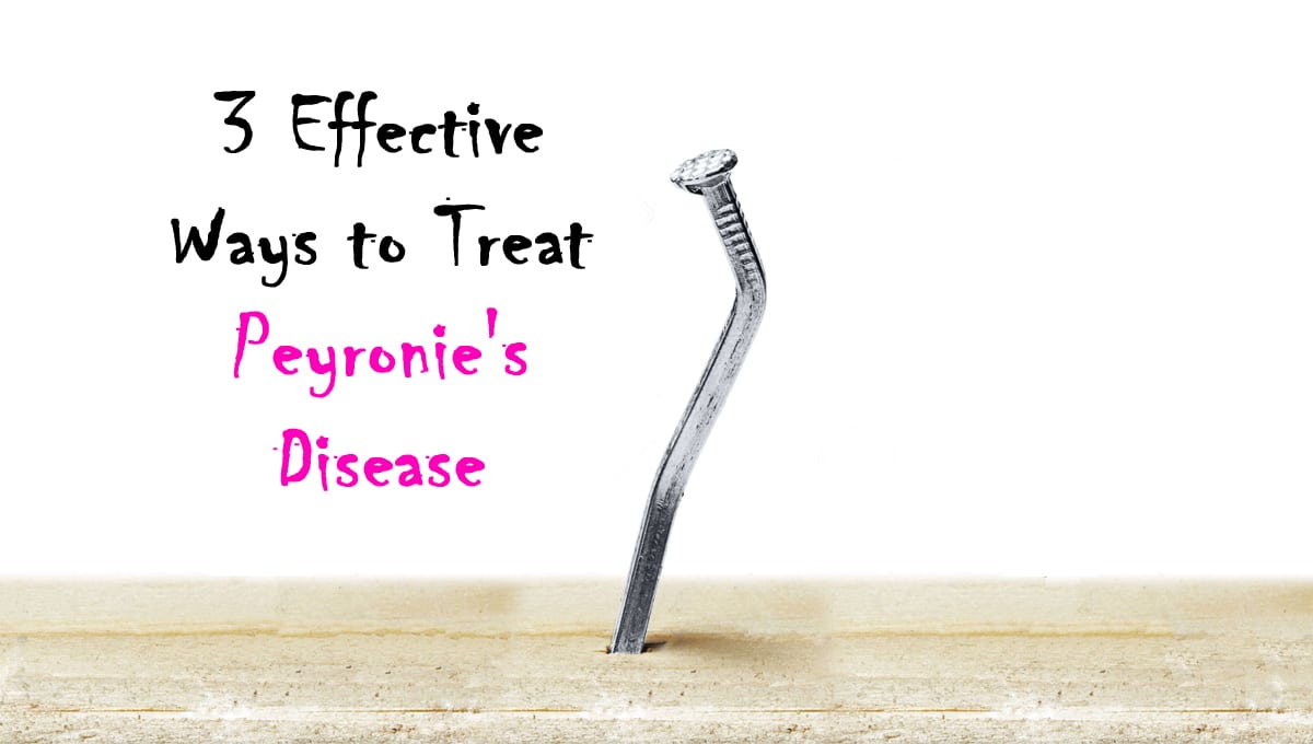
3 Effective Ways to Treat Peyronie’s Disease

Peyronie’s disease is a type of erectile dysfunction that leads to both physical and emotional scarring. It is caused by penile tissue damage resulting in the formation of scar tissue called plaque. Depending on the location and size of the plaque, the penis may bend downward or upward or become indented. The bending may occur suddenly (almost overnight) or happen gradually, beginning with soreness and lumps that develop into a hardened scar. While Peyronie’s disease is most often found in middle-aged men, it can occur at any age.
Here are 3 effective ways to treat Peyronie’s disease:

Medications

Drug therapy is helpful for most men who are affected by Peyronie’s disease, especially during the acute phase. For instance, oral Vitamin E will make the plaques smaller and help to straighten the bent penis. Potassium Amino-Benzoate (Potaba), Tamoxifen, PABA, Colchicine and Carnitine are also effective during the acute phase of the disease. Likewise, during the early stages of the condition, drugs for erectile dysfunction, such as sildenafil (Viagra), tadalafil (Cialis) and vardenafil (Levitra), may be beneficial.

As an alternative to oral medications, injecting certain drugs directly into the plaque can bring relief, particularly for men in the acute phase of the disorder who are not sure whether they want to undergo surgery. For example, verapamil injections disrupt the protein responsible for the formation of lesions in Peyronie’s disease, thus helping to relieve acute symptoms. Interferon injections prevent swelling and control scarring, while collagenase injections break down the plaque and are recommended for treating men with a penis curve of more than 30 degrees. Injection therapy may be administered using a combination of above drugs plus hyaluronidase, betamethasone or prostaglandin E1. Another method of applying these drugs is iontophoresis (electromotive administration/EMDA), where the drug is applied on the penile skin and then pushed through the skin using a low electric current.

Radiation Therapy

During radiation therapy, high-energy rays are targeted at the plaque to relieve the pain. The disadvantage of radiation therapy is that it relieves the pain on the bent penis without having a direct effect on the plaque which causes erectile dysfunction. Instead of radiation therapy, a man may opt for shockwave therapy, a form of mechanical treatment that uses a lithotripter device (such as those used for breaking up kidney stones) to break down calcified plaques or scarring. Other mechanical devices that can be used to treat the disease are low-dose radiotherapy and vacuum-based machines.

Surgery

Surgery for Peyronie’s disease is reserved for men suffering from severe, disabling penile disorder that makes it difficult to have sex. Before the surgery, the urologist will check the blood flow in the penis by injecting a drug to make it stiff and also examining the penis using ultrasound. The tests are used to assess the penis before deciding the type of surgery that is ideal. There are three basic ways through which the urologist will fix the disorder surgically. First, the urologist can remove the plaque and replace it with a tissue patch, helping to stretch the penis and restore its original length. Secondly, the surgeon may change or remove part of the tunica albuginea from the penis area opposite the plaque, an operation known as the Nesbit procedure. Lastly, the urologist may perform penile prosthesis implantation, a procedure recommended for men having both Peyronie’s disease and erectile dysfunction.

Conclusion

Since Peyronie’s disease varies extensively in how it affects patients, the treatment chosen should be carefully selected according to an individual’s condition. Most importantly, treatment should aim to preserve sexual function. If you are suffering from the disease, it is important to work closely with a urologist in order to get the right resources, prompt diagnosis and correct treatment, and to prevent the disease from turning into a crisis. For more information on dealing with Peyronie’s disease and erectile dysfunction, visit Advanced Urology Institute.

Effective Ways To Detect Prostate Cancer

If you are a man, there is a 1-in-6 chance that you will develop prostate cancer at some point in your life. Prostate cancer is basically a tumor of the prostate, the gland located in front of the rectum and just below the bladder. The prostate helps in making the milky fluid called semen, which carries sperm out of the body during ejaculation. Prostate cancer is most common among men over 65 years of age and fairly prevalent in men aged 50-64 years. Still, the cancer can occur in younger men below 50 and screening for it should begin between 40 and 45 years of age.

Risk factors of the cancer include:

  • (Age
  • Obesity
  • Family history
  • Race
  • History of STDs (sexually transmitted diseases)
  • Diets high in fats from red meat

While prostate cancer is a highly treatable condition, many men suffer needlessly because they did not know the symptoms in order to catch it before the advanced stages. And though the cancer typically shows few signs until it has reached advanced stages, knowing the symptoms can be the difference between having to face surgery and just making a few lifestyle changes. The symptoms of prostate cancer include:

In some instances of early prostate cancer, no symptoms occur and the cancer can only be detected through routine screenings.

Effective Ways to Detect Prostate Cancer

Your urologist will start by asking you about your medical history, symptoms and risk factors. A physical examination will also be performed. The doctor will then request a prostate-specific antigen (PSA) test to analyze your blood sample for higher-than-normal levels of the prostate-specific antigen, which is a protein secreted by the prostate gland. Higher PSA levels may indicate presence of cancer. In some cases, your doctor will opt to use the PSA3 test to check for levels of antigen 3 in your urine. When PSA levels are abnormal, the urologist will usually suggest that you return in 6 months for a second PSA test to confirm the presence of the cancer. Other test options that may be used to confirm a diagnosis of prostate cancer are:

(a) Digital rectal exam: The doctor will insert a gloved finger into your lower rectum and prostate gland to check for any abnormalities in size, texture or shape of the prostate.

(b) Targeted biopsy: The doctor removes tissue samples from the prostate by using either MRI fusion-targeted 3D imaging method or the ultrasound high-yield saturation method. Samples taken are analyzed to detect the cancer and determine its severity.

(c) Prostate imaging: Also called perfusion dynamic MRI, prostate imaging uses a special contrast agent inserted into the prostate to precisely identify patients with the risk of prostate carcinoma.
Once prostate cancer is confirmed, the next step is determining its aggressiveness. The aggressiveness of a cancer helps the urologist to choose the best treatment option for the condition. So, after knowing how aggressive the cancer is, the urologist can recommend:

(a) Active surveillance: You are not offered any treatment but kept under careful observation and medical monitoring.

(b) External beam therapy (EBT): The urologist refers you to a radiation oncologist to deliver a beam of high-energy proton beams or x-ray to the tumor location. The oncologist may also use other radiation techniques such as conformal external beam radiation therapy, stereotactic body radiation therapy, proton beam therapy, or image-guided radiation therapy.

(c) Brachytherapy: In this case, radiation is delivered to your prostate by placing radioactive materials inside the prostate. The two forms used are low-dose rate (LDR) and the high-dose rate (HDR) Brachytherapy.

(d) Cryotherapy: This method uses extremely low temperatures (as low as -190 degrees Celsius) to freeze and destroy cancer cells.

(e) Surgery: The urologist makes an incision through the perineum or in the lower abdomen to remove the prostate. A skilled and experienced urologist will perform the surgery without interfering with your sexual function.
At Advanced Urology Institute, we have the best pool of urologists and state-of-the art equipment to help detect and treat prostate cancer. If you want to be screened for the condition, even without symptoms, never hesitate to see a urologist. Remember, early detection is the key to successful prostate cancer treatment. To learn more about prostate cancer screening, prevention and treatment, visit Advanced Urology Institute.

Why Walnuts Make Men More Fertile

Are you a man struggling to have children and looking for a way to boost your fertility? If so, consider eating a handful of walnuts every day. According to a study conducted by the University of Delaware, eating a walnut-enriched diet boosts sperm quality and aids in preventing male infertility. Working with two groups of mice, one fed on a walnut-enriched diet and the other fed on a control diet for 9 weeks, the researchers found that the group fed walnuts had a significant improvement in sperm quality.

How Do Walnuts Work?

Walnuts reduce lipid peroxidation, a form of cell damage that interferes with sperm membranes and harms sperm cells. Sperm is made of polyunsaturated fatty acids which are liable to damage by lipid peroxidation. Walnuts are the only tree nut made of mostly polyunsaturated fatty acids and will therefore effectively replenish sperm cells. In fact, just 1 ounce of walnuts contains 13 grams of polyunsaturated fatty acids (PUFAs) in 18 grams of total fat. By preventing sperm damage and replenishing sperm cells, walnuts help to improve sperm morphology, motility and vitality.

First Study on Walnut Efficacy

The study by the University of Delaware was based on a previous study by the UCLA Fielding School of Public Health and School of Nursing. According to the first study, which was based on a randomized control trial at UCLA, eating 2.5 ounces of walnuts each day (about 30 walnut halves) significantly improves the motility, morphology and vitality of sperm in men. During the research, more than 100 healthy young men eating their usual Western-style diet participated in monthly calls to share what they ate throughout the study. At the end of the study, it was clear that walnuts improved sperm quality, though more research was necessary to verify the role of the nuts.

Second Research Study by the University of Delaware

When the University of Delaware conducted its study, it was designed to reveal the mechanism by which walnuts had improved sperm quality in the UCLA study. Two groups of male mice, one consisting of healthy male mice and the other of genetically predetermined infertile male mice, were fed on either a walnut-enriched diet or a controlled diet for 9 to 11 weeks. At the end of the study, significant improvements in sperm morphology and motility were observed in mice consuming 20% of their daily calories from walnuts (equivalent to 2.5 ounces daily in humans). Most interestingly, even infertile mice had a remarkable boost in sperm morphology and both groups showed noteworthy reduction in peroxidative damage. The researchers concluded that walnuts improve sperm quality by reducing peroxidative sperm cell damage, though recommending further studies to determine which specific nutrients in walnuts are responsible for this improvement.

Walnuts are Beneficial for Sperm Health

Fertility in men is determined largely by sperm quality and quantity. For instance, if the number of sperm ejaculated is very low or the sperm are of poor quality, it may be difficult or even unlikely for pregnancy to occur. Since one-in-five infertility cases are solely due to the male partner and about one-in-twenty men suffer from some form of fertility problem related to low numbers of sperm ejaculated, using a healthy and proven method of improving sperm morphology, vitality and motility is a good way to begin addressing fertility problems. Enriching your diet with walnuts can be an effective first step in resolving your fertility issues and, if needed, there are many other treatments for infertility using medications or surgery. For more information on dealing with male infertility, visit Advanced Urology Institute.

5 Effective Treatment Options for Erectile Dysfunction

5 Effective Treatment Options for Erectile Dysfunction

There are several treatment options available for erectile dysfunction (ED), and the right one for an individual depends on the cause of the ED, age, health and lifestyle of the patient. In most cases, the use of oral medications such as Cialis, Viagra or Levitra is the recommended first option. Then, depending on the effectiveness and tolerance of the medication, other treatments may be tried. For example, microvascular surgery is recommended for patients with blood vessel leakage or blockage, while penile prosthesis is one effective alternative for those who fail to respond to non-surgical therapy or are not candidates for surgery.
Here is a look at 5 effective treatment options for erectile dysfunction:

1. Oral Medications: Phosphodiesterase-5 (PDE-5) inhibitors

Phosphodiesterase-5 (PDE-5) inhibitors, such as sildenafil (Viagra), tadalafil (Cialis), vardenafil (Levitra) and avanafil (Spedra), are some of the most widely used and highly effective medications for treating erectile dysfunction. The pills work by temporarily boosting the flow of blood to the penis. Sildenafil, sold as Viagra by Pfizer, is the most commonly prescribed pharmacological medicine for ED. Though it is available in different forms, Viagra takes 30-60 minutes to show effect and has a 4-hour duration. The drug only boosts blood flow to the penis and therefore sexual arousal and stimulation are necessary in order to have and maintain an erection. Tadalafil (Cialis) has effects lasting longer (up to 36 hours depending on the dose taken) and takes effect sooner (15-30 minutes). Tadalafil is also more selective in action and does not show the side effects of sildenafil and vardenafil. Avanafil (Spedra) and vardenafil (Levitra) show similar mechanism of action as sildenafil (Viagra) and only differ in duration of onset and length of effect.

2. Urethral Suppository (MUSE)

The use of a medicated urethral system for erection (MUSE) is considered when oral pills are not effective. The treatment involves placing a small medicated suppository in the penile urethra without using needles so that the suppository is absorbed to help produce an erection. The main advantage of this treatment option is that the suppository is applied locally by the patient or his partner, and has very few side effects. After the suppository is inserted into the urethra, sexual stimulation is necessary for increased blood flow to the penis. Patients opting for MUSE should have the first suppository application done in the urologist’s office to prevent potential complications such as decreased blood pressure, urethral bleeding, and continued and prolonged erections.

3. Penile Injections

The urologist may recommend penile injections when oral medications prove ineffective. And even though the idea of injecting the penis may be quite unappealing, the effectiveness and ease of injections make this a worthwhile option to consider. Apart from Caverject and Edex that have been prescribed by urologists for a while, other commonly injected medications are papaverine, phentolamine and alprosdid. Often, urologists recommend a blend of two or three medications for use in the injections. The blend, called a Trimix, ensures a synergistic effect of three medications, keeps the dose of every drug low to prevent adverse effects, and delivers a response rate of up to 90%.

4. Vacuum Pumps

Vacuum devices are recommended for patients who have only partial erections and find other treatments ineffective or intolerable. A vacuum device is made of a plastic cylinder connected to a pump and a constriction ring. Using a battery power or manual pressure to create suction around the penis in order to bring blood into it, a vacuum pump causes an erection which is maintained by a tourniquet of sorts that is placed around the base of the penis and an elastic rind which stops additional blood from flowing out of the engorged penis. Vacuum pumps are a relatively inexpensive, safe and easy to use treatment option for erectile dysfunction with minimal side effects. Erections induced using pumps generally last for about 30 minutes.

5. Penile Prosthesis and Penile Vascular Reconstructive Surgery

Surgery is considered as the last resort when all other treatment options fail. However, it may be the best option for young men seriously injured in their pelvic area (such as in a car accident) and men with significant anatomical problems with their penis. A penile prosthesis is an effective and more invasive option in which either a semi-rigid or inflatable implant is placed on the penis through surgery. The use of penile prosthesis has been proven to offer up to 85% patient-partner satisfaction rate. Penile vascular (venous or arterial) surgery is recommended for young men who have erectile dysfunction due to congenital or traumatic leakage of the penis. Penile venous surgery is performed to boost the trapping of blood in the penis, boosting a man’s capacity to get and maintain erections. On the other hand, penile arterial surgery creates a path of blood flow to the penis by correcting/bypassing blocked arteries.

For more information on effective treatments of erectile dysfunction, visit the Advanced Urology Institute website or make an appointment with a urologist today.

3 Easy Ways to Prevent Kidney Stones

3 Easy Ways to Prevent Kidney Stones


Kidney stones are formed when certain chemicals present in the urine solidify and turn into hard crystals. Over time, these crystals grow in size and eventually leave the body through urine. Sometimes, the crystals get stuck in the urinary tract, blocking the flow of urine and causing enormous pain. In nearly 50% of patients with this problem, the stones reappear within 5 to 7 years if no preventive measures are taken. In most cases, these stones form when calcium reacts with phosphorus or oxalate. A physician first determines the cause of this condition and will likely suggest reducing the intake of sodium or protein, both of which cause kidney stone formation. With some determination and care, the risk of kidney stones can be significantly reduced. The following are three simple preventive methods:

1. Increase Calcium

Calcium deficiency causes an increase in the body’s oxalate levels which directly contributes to the formation of stones. Find out how much calcium you should consume for your age to ensure that your body is not deficient in this essential mineral. Generally, men older than 50 years of age need 1,000 mg of calcium every day in addition to 800 to 1,000 IU of vitamin D to help the body absorb calcium. Getting your calcium from food is preferable since studies show that calcium supplements may increase the risk of stone formation.

2. Reduce Animal Protein

Your body produces uric acid while breaking down proteins. Higher levels of this compound increase the acidity of urine, which may cause the formation of kidney stones in the long run. For this reason, keep a check on protein-rich foods, especially red meat, seafood, poultry, and eggs. Eating too much protein also reduces the content of citrate in your urine, and this may also lead to stone formation. If you are prone to kidney stones, you should also follow a low-sodium meal plan. Nutritionists suggest a daily maximum sodium intake of 2,300 mg, but people who already have kidney stones due to high sodium levels should consume less than 1,500 mg of sodium per day.

3. Drink Plenty of Fluids

Drinking plenty of water is the simplest home remedy for kidney stones because water dissolves the unwanted substances in urine. As a rule of thumb, drink at least 2 liters of water per day. You may substitute with citrus beverages such as fresh orange juice or lemonade.

In addition to the above measures, avoid stone-forming foods such as chocolate, beets, tea, nuts, rhubarb and spinach, all of which are rich in oxalate. Colas are high in phosphate, a substance to avoid if you have a history of kidney stones due to high phosphate levels. Also, our bodies turn vitamin C into oxalate, so individuals taking this vitamin in supplement form can be at a slightly higher risk. Overall, with proper treatment and some changes to your diet, kidney stones can easily be prevented.

7 Types of Bladder Control Problems in Women

Bladder control problems are common for many women. They are characterized by urine leakage, a progressively weaker urine stream, inability to empty the bladder or the frequent urge to urinate and rush to a bathroom. Bladder issues often restrict a woman’s range of physical activities and may cause withdrawal from social interactions, resulting in a diminished quality of life. Causes may include weakness of the pelvic fascio-muscular supports, nerve damage, various medications for neurologic conditions and underlying medical disorders like kidney disease and diabetes. While these problems differ according to cause and contributing factors, urologists can help women regain bladder control by identifying the type of bladder problem and administering the right treatment.
The 7 most common types of bladder control problems in women are:

1. Temporary or transient incontinence

This is a short-lived episode of loss of bladder control that commonly affects more than half of hospitalized women and at least a third of community-dwelling elderly women. It is often a side effect of medications (such as sleeping pills and diuretics) that lower cortical control over the urinary bladder or stimulate overproduction of urine. They may also be caused by surgery, pregnancy, urinary infections, severe constipation, or an inflamed or irritated bladder, vagina or urethra. In all cases, temporary incontinence will resolve on its own as soon as causative factors are identified and corrected.

2. Stress incontinence

This is the involuntary loss of bladder control associated with activities that increase physical pressure and stress in the abdomen and bladder. Affected women report having urine escapes when they laugh, cough, sneeze, have sex, exercise or engage in heavy lifting. It is the most common type of incontinence among women, occurring at any stage of life due to the physical changes of pregnancy, childbirth or menopause, all of which weaken the pelvic floor and reduce the effectiveness of bladder supporting ligaments.

3.Urge incontinence

Urge incontinence is the loss of bladder control commonly resulting from abnormal nerve signals or nerve damage due to a cerebrovascular accident, an infection or diabetes mellitus. It is characterized by a strong, abrupt and urgent need to urinate without prior warning, followed by the escape of a considerable amount of urine almost at once. Women affected by this bladder problem report leaking urine on their way to the restroom, urinate more than eight times per day and usually have to get to the bathroom more than two times overnight.

4. Mixed incontinence

This type of incontinence represents a combination of the characteristics of both urge and stress incontinence, with affected women experiencing episodes of sudden, urgent and uncontrollable urge to urinate together with urine leakage after a sudden cough, sneeze or laughter. All the factors that trigger urge and stress incontinence also cause mixed incontinence, including abnormal nerve signals, nerve damage, weakened pelvic floor muscles and connective tissue abnormalities.

5. Functional incontinence

Unlike the other types of incontinence, functional incontinence is not caused by abnormalities in the urinary system, nerves or muscles, but it occurs when a woman is unable to reach the bathroom in time to void because of mental and physical limitations. For example, a woman who is handicapped (such as having arthritis, a broken leg, Parkinson’s disease or Alzheimer’s disease) may not be able to move to the bathroom to urinate as soon as her bladder capacity is far exceeded, urinating where she is. Therefore, functional incontinence is a side effect of a mental or medical health issue.

6. Overflow incontinence

Characterized by the involuntary leakage of small amounts of urine when the amount of urine in the bladder exceeds the bladder’s maximum capacity, overflow incontinence occurs in women with weak bladder muscles, blocked urethra, kidney stones, scar tissue, pelvic organ prolapse, diabetes and multiple sclerosis. In overflow incontinence, the bladder has a substantial amount of residual urine and tends to overfill rapidly, resulting in the overflow of urine within a very short time. When not promptly treated, overflow incontinence can lead to bladder infection.

7. Overactive bladder

In some women, the inability to control the bladder is characterized by a sudden and unstoppable need to urinate, passing urine eight or more times per day and having to wake up two or more times every night to pass urine. In women with overactive bladder, detrusor muscles contract unpredictably and the inability to control the bladder can be so embarrassing that the affected person may want to isolate herself and limit her work and social life.
For women having bladder control problems, the good news is that a brief evaluation by an experienced urologist can quickly identify the type of bladder problem and pave the way for immediate treatment. If you have bladder control problems, inform your doctor and ask for help. Do not let your embarrassment keep you from getting the help you need. For more information, visit the site Advanced Urology Institute.

Why I Love Urology! Treating and Curing Urological Conditions

Why I Love Urology! Treating and Curing Urological Conditions

Urology is a medical field that deals in the diagnosis, treatment, curing and management of conditions that affect the male and female urinary system and the male reproductive organs. It is a dedicated branch of medicine and the physicians who are specialists in this field are called urologists.The medical disorders treated by urologists include those that affect the kidneys, ureters, bladder, urethra, prostate, epididymis, testis, seminal vesicle, scrotum and penis. Common conditions include urinary incontinence, ejaculation problems, erectile dysfunction, vasectomy, cryptorchidism (undescended testes), vesicoureteral reflux and genitourinary cancers such as prostate cancer, cancer of the kidneys, bladder, penis and testicles.

Urologists love their jobs because it gives them opportunities to provide patients with life saving treatments as well as quality of life medical care. Conditions such as cancer of the testes, if diagnosed early, can be treated to give the patient a longer life span and one free of discomfort or pain during intimacy.

It is important to note that the work of a urologist also involves educating the general public on effective prevention practices. A good example is prostate cancer, an illness which has been on the rise recently. Research has identified potential causes, prevention and treatment. This knowledge has been shared widely by urologists. Men are now encouraged to use testing for early diagnosis. If caught in the primary stages, prostate cancer can be cured before it progresses to stage IV cancer or metastasizes to other parts of the body. The same applies to other conditions such as erectile dysfunction, a topic most men prefer to avoid. Despite the reluctance to acknowledge this medical problem, in recent years the public has been receiving education from the experts on the many causes and treatments of erectile dysfunction.

Urology is a field that specializes in treating urological physical ailments and the emotional concerns that accompany them. Each patient presents with different needs and urologists who love the work they do understand that. They take into account the presenting factors of each patient, their needs and concerns, and then tailor the consultations, medical treatment and follow up to suit the unique needs of each patient. Any person with concerns about a potential urological problem should pay a visit to a qualified specialist for a check-up. The experienced and caring professionals of the Advanced Urology Institute would like to remind us that the earlier a problem is diagnosed, the more successful the treatment will be.

Why do I have a curved penis, Could it be a symptom of Peyronie’s disease?

The penis tends to bend slightly when it is erect because human anatomy is rarely perfectly symmetrical. The direction of bending of an erect penis depends on the ratio of the crus (portion of penis under the skin) to the exposed penis. Hence, a man with a shorter crus and longer exposed penis tends to have an erection pointing downward while one with a longer crus and shorter exposed penis has an erect penis pointing straight up or outward. In some men, the penis may curve slightly to the right or to the left. However, when the bend is more extreme and accompanied by painful erections, it may be a symptom of Peyronie’s disease.

What Is Peyronie’s Disease?

Peyronie’s disease (PD) is the most common cause of a curved penis. As a collagen disorder characterized by scar tissue (plaques) that develops under the skin, Peyronie’s disease is typified by a bend of the penis during erections. Other deformities, such as penis shrinkage, loss of girth, hourglass narrowing and indentation may also occur. The resulting bend makes penetrative sex painful or difficult, culminating in physical and psychological distress that causes difficulties in relationships.While the actual cause of Peyronie’s disease is not known, genetic defects, damage to blood vessels of the penis and low levels of testosterone hormone are believed to cause the disorder. And although the number of men who develop the condition is not known—given that most men are too embarrassed to see a doctor about the disorder—it is estimated that between 3-9 men out of 100 are affected. The disease is common among men in their fifties, though a small number of teenagers may also develop it.

Diagnosis and Treatment of Peyronie’s Disease

Typical symptoms such as painful erections, bending, deformity of the shaft and angulation are indicative of the disease. Given these indications, the urologist will measure the bend or distortion of an erect penis using photographs taken by a patient at home, a vacuum pump, or an injection to stimulate an erection. In some cases, the urologist may find it necessary to order a duplex ultrasound test in order to have a more accurate assessment of blood circulation in the penis.

Treatment options for the disease include stretching (external penile traction), vacuum devices, oral medicines (such as para-aminobenzoate, propoleum and colchicines), topical medicines (applied on the surface of the penis, like topical verapamil), injections into scar tissue (such as interferon, verapamil and clostridium hystolyticum (Xiaflex)), electrical currents (iontophoresis) and surgery.

So, is it normal to have a curved penis or could it be a symptom of Peyronie’s disease? Well, a mild curvature may be normal, but a severe curvature accompanied by painful erections requires immediate medical attention as it may, in fact, be a symptom of a curable disease. Presently, urologists have several treatment options for handling the condition in a personalized manner. Don’t suffer in silence. For more information, visit Advanced Urology Institute.

Life Changing Treatment At Advanced Urology Institute

What I Do as a Physician Assistant – Lisa Cunningham


Driven by an unwavering commitment to excellence in urology, Advanced Urology Institute offers a complete range of innovative, specialized and high quality medical services for male and female urological conditions. We have assembled a multidisciplinary team of specialists who work together for you, including preoperative nurses, urologists, pathologists, radiation oncologists, dedicated robotic surgeons, postoperative recovery specialists and nurse navigators who coordinate urologic cancer care. Our board certified experts will work with you to identify the most effective treatment that meets your individual needs, ensuring a minimal disruption of your lifestyle.

Better Outcomes

At Advanced Urology Institute, we recognize that the most effective treatment for urologic conditions requires close collaboration between urologists, oncologists and other medical specialists. We have therefore created an environment in which the interactions between different specialists result in better patient care and improved outcomes. Our specialists work together managing each case jointly right from the start because we have removed all artificial boundaries between specialties, thus ensuring that transitions are seamless. In fact, we can deliver comprehensive treatment from one location by combining surgical expertise, on-site pathology and clinical laboratories, imaging services such as radiation therapy and CT scan, leading-edge robotic technology and synchronized care, resulting in unmatched personalized patient experience.

Comfort and Convenience

As experts on the conditions of the female and male urinary tract and the male reproductive system, we endeavor to help patients navigate sensitive health issues with a focus on their comfort and convenience. Every urologist at our center is open, friendly and willing to listen to the unique story of each patient in order to correctly recognize the symptoms and provide proper diagnosis. This extends to the use of the most recent diagnostic tools and the development of personalized treatment programs for conditions such as kidney cancer, bladder cancer, prostate cancer, male erectile dysfunction, renal pelvic obstructions, adrenal disease and female urologic disorders.

Are you looking for the highest quality urology services? Visit our site ‘Advanced Urology Institute‘ for more information.

What Are The Types of Hematuria?

Video: Providing Effective Patient Education

Hematuria is a health condition that is characterized by the presence of blood in the urine. There are two main types of hematuria: microscopic and macroscopic.

In microscopic hematuria, there is no visual evidence of the presence of blood, unless the urine is view under a microscope. Macroscopic hematuria, on the other hand, is evident immediately with red discoloration of the urine.

Hematuria can also be classified according to the cause of the blood in the urine. Both of these types are detailed further below.

Microscopic Hematuria

The concentration of blood in the urine is not visible to the naked eye in microscopic hematuria and can only be detected under a microscope.

Some individuals are affected by microscopic hematuria without an identifiable cause, which is referred to as idiopathic hematuria. This is thought to result from an increased excretion of red blood cells in respect to what is considered to be normal, although may not be associated with adverse effects on the individual.

Macroscopic Hematuria

Also known as frank or gross hematuria, macroscopic hematuria involves visible discoloration of the urine as a result of a greater concentration of blood in the urine. The color is usually described as pink, red or dark brown. Additionally, there may also be evidence of small or large blood clots in some cases.

It is not necessary for a large volume of blood to be present for the color of the urine to be altered. In fact, 1 mL of blood is sufficient to precipitate a change in color. Additionally, the volume of blood may not be an accurate indicator of the severity of the underlying cause, and even a small quantity of blood may have serious repercussion and vice versa.

Joggers Hematuria

“Joggers hematuria” is a specific type of hematuria that occurs as a result of recurrent damage to the bladder during activities such as jogging and long-distance running.

Classification by Cause

Hematuria can also be classified according to the cause of the condition, as follows:

  • Infective hematuria: due to pyelonephritis, cystitis or urethritis
  • Stones-related hematuria: due to staghorn calculi, calcium stones or uric acid stones
  • Trauma-related hematuria: due to pelvic trauma, renal injuries or foreign bodies
  • Renal hematuria: due to IgA nephropathy, hereditary nephritis, medullary sponge kidney or thin basement membrane diseases
  • Iatrogenic hematuria: due to recent endoscopic procedure, trans-rectal ultrasound, traumatic catheterization, radiation, indwelling ureteric stents, renal biopsy or extracorporeal shockwave lithotripsy
  • Benign hematuria: due to strictures, renal masses or benign prostatic hypertrophy
  • Malignant hematuria: due to prostate acinar adenocarcinoma or renal cell, transitional cell, squamous cell or urothelial cell carcinoma

Other Causes of Red Discoloration

While red discoloration is the most distinctive feature of hematuria, there are various other causes of this discoloration, which can result from numerous factors. These may include the presence of pigments such as:

  • Myoglobin (indicative of myoglobinuria)
  • Porphyrins (indicative of porphyria)
  • Betanin (in beets)

Additionally, some drugs can have a similar effect, including rifampicin, phenazopyridine, sulphonamides and non-steroidal anti-inflammatory drugs (NSAIDs).

[Read Original Article…]